• Biochemistry and Molecular Biology
  • Biostatistics
  • Environmental Health and Engineering
  • Epidemiology
  • Health Policy and Management
  • Health, Behavior and Society
  • International Health
  • Mental Health
  • Molecular Microbiology and Immunology
  • Population, Family and Reproductive Health
  • Program Finder
  • Admissions Services
  • Course Directory
  • Academic Calendar
  • Hybrid Campus
  • Lecture Series
  • Convocation
  • Strategy and Development
  • Implementation and Impact
  • Integrity and Oversight
  • In the School
  • In the Field
  • In Baltimore
  • Resources for Practitioners
  • Articles & News Releases
  • In The News
  • Statements & Announcements
  • At a Glance
  • Student Life
  • Strategic Priorities
  • Inclusion, Diversity, Anti-Racism, and Equity (IDARE)
  • What is Public Health?

Is the Hygiene Hypothesis True?

Did Covid shutdowns stunt kids' immune systems?

Caitlin Rivers

The hygiene hypothesis is the idea that kids need to be exposed to germs in order to develop healthy immune systems. We know that many common viruses did not circulate as widely during the pandemic, thanks to social distancing, masking, and other COVID mitigation measures. Are there downsides to those missed infections? 

In this Q&A, Caitlin Rivers speaks with Marsha Wills-Karp, PhD, MHS , professor and chair of Environmental Health and Engineering , about the role of household microbiomes, birth, and vaccines in the development of kids’ immune systems—and whether early exposure really is the best medicine.

This Q&A is adapted from Rivers’ Substack blog, Force of Infection .

I think there’s some concern among parents who have heard about the hygiene hypothesis that there is a downside to all those stuffy noses that didn’t happen [during the COVID-19 pandemic]. Are there any upsides to viral infections? Do they help the immune system in some meaningful way?

I don’t think so.

You mentioned the hygiene hypothesis, which was postulated back in the ‘80s. German scientists noticed that families with fewer children tended to have more allergic disease. This was interpreted [to mean] that allergic disease was linked to experiencing fewer infections. I have explored this idea in my research for a couple of decades now.

This phenomenon has helped us to understand the immune system, but our interpretation of it has grown and expanded—particularly with respect to viruses. Almost no virus is protective against allergic disease or other immune diseases. In fact, infections with viruses mostly either contribute to the development of those diseases or worsen them.

The opposite is true of bacteria. There are good bacteria and there are bad bacteria. The good bacteria we call commensals . Our bodies actually have more bacterial cells than human cells. What we’ve learned over the years is that the association with family life and the environment probably has more to do with the microbiome. So one thing I would say is sanitizing every surface in your home to an extreme is probably not a good thing. Our research team showed in animals that sterile environments don’t allow the immune system to develop at all. We don’t want that.

What does contribute to the development of the immune system, if not exposure to viruses?

There are a number of factors that we’ve associated with the hygiene hypothesis over the last 20 years, and these exposures start very early in life. Cesarean sections, which do not allow the baby to travel through the birth canal and get exposed to the mother’s really healthy bacterial content, is a risk factor for many different immune diseases. Getting that early seeding with good bacteria is critical for setting up the child going forward. Breastfeeding also contributes to the development of a healthy immune system.

There are other factors. Our diets have changed dramatically over the years. We eat a lot of processed food that doesn’t have the normal components of a healthy microbiome, like fiber. These healthy bacteria in our gut need that fiber to maintain themselves. They not only are important for our immune system but they’re absolutely critical to us deriving calories and nutrients from our food. All these things contribute to a healthy child.

We’ve also noticed that people who live on farms have fewer of these diseases because they’re exposed to—for lack of a better term—the fecal material of animals. And what we have found is that it’s due to these commensal bacteria. That is one of the components that help us keep a healthy immune system. Most of us will probably not adopt farm life. But we can have a pet, we can have a dog.

I think all the pet lovers out there will be pleased to hear that.

There’s a lot of evidence that owning a pet in early childhood is very protective.

What about the idea that you need to be exposed to viruses in early life because if you get them as an adult, you’ll get more severely ill? We know that’s true for chickenpox, for example. Do you have any concerns about that?

We should rely on vaccines for those exposures because we can never predict who is going to be susceptible to severe illness, even in early childhood. If we look back before vaccines, children under 4 often succumbed to infections. I don’t think we want to return to that time in history.

Let me just give you one example. There’s a virus called RSV, it’s a respiratory virus. Almost all infants are positive for it by the age of 2. But those who get severe disease are more likely to develop allergic disease and other problems. So this idea that we must become infected with a pathogenic virus to be healthy is not a good one.

Even rhinovirus, which is the common cold, most people recover fine. But there’s a lot of evidence that for somebody who is allergic, rhinovirus exposures make them much worse. In fact, most allergic or asthmatic kids suffer through the winter months when these viruses are more common.

And that’s particularly salient because there is a lot of rhinovirus and enterovirus circulating right now.

From my point of view, right now, avoiding flu and COVID-19 is a priority. Those are not going to help you develop a healthy immune response, and in fact, they can do a lot of damage to the lungs during that critical developmental time. Data [show] that children that have more infections in the first 6 months to a year of life go on to have more problems.

It’s always surprising to me when I look at the data of the fraction of time that young children spend with these common colds—and this is pre-pandemic—it’s not uncommon for kids to be sick 50% of the time. That feels right as a parent, but it’s startling.

The other thing people don’t know is that the GI tract is where you get tolerized to all of your foods, allergens and things. Without those healthy bacteria in your gut, you can’t tolerate common allergens.

How does that relate to the guidance that’s changed over the years—that you should withhold peanuts in early life and now you’re supposed to offer them in early life?

The guidance to delay exposure to peanuts didn’t consider the fact that oral exposure to peanuts was not the only exposure kids were getting. There were peanut oils in all kinds of skin creams and other things. So kids got exposed through their skin, but they had no gut protection—and the GI tract is important for a tolerant system. If you have a healthy immune response, you get tolerized in early life.

This concept is a little bit different for those families who may already have a predisposition to allergies. But for the general public, exposure is key to protecting them in early life.

I think some parents look at the guidance that you should now offer peanuts in early life and say, “Are we not doing that with rhinovirus by masking kids or improving ventilation?” How should people think about the development of the immune system for food allergies compared to infections?

The thing about rhinoviruses is that after recovering, you’re not protected from the next infection. There is no real immune protection there. Most of us suffer from colds throughout our whole life. Like I said, bacterial exposure is what’s key to priming the immune response. 

Also, we forget that a lot of kids die from the flu. Unlike COVID-19, where younger kids are not quite as susceptible to severe illness, that’s not true for flu. RSV, too, can be quite severe in young children and older adults.

Caitlin Rivers, PhD, MPH , is a senior scholar at the Johns Hopkins Center for Health Security and an assistant professor in Environmental Health and Engineering at the Johns Hopkins Bloomberg School of Public Health.

RELATED: 

  • Study Finds That Children’s Antibody Responses to COVID-19 Are Stronger Than Adults’
  • Back to School: COVID, CDC Guidance, Monkeypox, and More
  • A New Shot Prevents Serious Illness from RSV

Related Content

A vehicle is left abandoned in floodwater on a highway after Hurricane Beryl swept through the area on July 8, 2024, in Houston, Texas.

The Visible and Unseen Dangers Lurking in Floodwater

Commuters walk under the Olympic rings at the Gare de Lyon train station, on July 23, 2024, ahead of the Paris 2024 Olympic and Paralympic Games.

Public Health Prep for the 2024 Paris Olympics

A nurse assists a patient on a hospital bed with a pulse oximeter

The Problem with Pulse Oximeters: A Long History of Racial Bias

Illustration of red blood cells affected by sickle cell disease.

How Sickle Cell Disease and Malaria Defined Evolution

Tikur Anbessa Specialized Hospital, Addis Ababa University medical campus on sunny day with a blue sky background.

A Forgotten, Yet Life-Threatening Infection

The hygiene hypothesis: How being too clean might be making us sick

by Joseph Stromberg

what is a hypothesis for bacteria

Over the past few decades, doctors have arrived at a counterintuitive hypothesis about our modern, ultra-sanitized world. Too much cleanliness may be causing us to develop allergies, asthma, inflammatory bowel diseases, and other autoimmune disorders.

The idea is that for many children in the wealthy world, a lack of exposure to bacteria, viruses, and allergens prevents the normal development of the immune system, ultimately increasing the chance of disorders within this system down the road. This is called the hygiene hypothesis .

a lack of exposure to bacteria, viruses, and allergens may prevent the normal development of the immune system

“A child’s immune system needs education, just like any other growing organ in the human body,” says Erika von Mutius, a pediatric allergist at the University of Munich and one of the first doctors to research the idea. “The hygiene hypothesis suggests that early life exposure to microbes helps in the education of an infant’s developing immune system.” Without this education, your immune system may be more prone to attacking the wrong target — in the case of autoimmune diseases, yourself.

It's still a matter of active debate among scientists, but e vidence for the idea has been slowly accumulating over time, both in humans and animal subjects. It's been cited as an explanation for why allergy and asthma rates are so much higher in wealthy countries, and m ost recently, a study published last year found that babies who grow up in houses with higher levels of certain bacteria — carried on cockroach, mouse, and cat dander — are less likely to develop wheezing and asthma by the age of three.

(However, it’s important to note that despite the claims of some anti-vaccine activists, there’s absolutely no evidence that not getting vaccinated has similar benefits.)

How could this kind of filth possibly make us healthier? Here’s an explanation of the hygiene hypothesis.

How doctors got the idea that dirt could make us healthy

141479142

(REMY GABALDA/AFP/Getty Images)

Obviously, the basic sanitary practices we've developed as a society over the past few centuries — such as building infrastructure to remove garbage and sewage from cities — have provided all sorts of benefits. They're a huge part of the reason so few Americans get infectious diseases like cholera or typhoid nowadays.

asthma, hay fever, and other allergies have become much more common as we've become more sanitary

But researchers have found that a few specific autoimmune diseases — asthma, hay fever , inflammatory bowel diseases, and various allergies — have become much more common as we've become more sanitary, and are much more prevalent in the wealthy world than the developing one.

In the late 1980s, when studying childhood allergies in East and West Germany, British epidemiologist David Strachan began to suspect there was a connection. In the dirtier, more polluted, less wealthy cities of East Germany, he found, children had much lower rates of hay fever and asthma than in the cleaner, richer cities of West Germany.

To explain this, he looked at all sorts of lifestyle differences — and found that West German children were much less likely to spend time in day care centers, around other kids, than East German children. He proposed that their reduced exposure to bacteria and other antigens, normally acquired from other children, somehow affected their immune systems, leading to their increased chance of developing the autoimmune diseases.

The evidence for the hygiene hypothesis

183666877

Children who grow up on farms have lower rates of allergies. (John Moore/Getty Images)

In the decades since, all sorts of epidemiological evidence has been collected that supports Strachan’s idea. He initially found that in Britain, children who grew up in larger families also had lower chances of developing asthma and hay fever , presumably because they were exposed to more bacteria from their siblings.

Other doctors have found that, on the whole, people in wealthy, more heavily sanitized nations have much higher rates of asthma and allergies than those in the developing world. This could be a function of natural variations among the populations, but more recently, doctors have found that people who move from a developing country to a wealthier one have a higher chance of developing these diseases than people who stay in their country of origin.

kids who grow up on farms or have pets have lower rates of allergies and asthma

Even within a developing country like Ghana, wealthy urban children have higher rates of these autoimmune diseases than poorer or rural children. In the wealthy world, adults who clean their houses with antibacterial sprays have higher asthma rates, and people who are more often exposed to triclosan (the active ingredient in antibacterial soap) have higher rates of allergies and hay fever . Kids who grow up on farms or have pets, meanwhile, have lower rates of allergies and asthma .

These are all correlations — not causations — but they suggest that something about the relatively clean, modern urban environment makes these autoimmune diseases more likely to develop. And the handful of controlled studies conducted on the topic have provided further support — such as one, conducted recently in Uganda , in which babies born to mothers who were given drugs to treat parasitic worm infections during pregnancy ended up having higher rates of eczema and asthma.

Controlled studies with animals have also provided compelling evidence for the idea. “In experimental studies with germ-free mice raised in a sterile environment, researchers have found they’re extremely prone to developing colitis and asthma, among many other problems,” von Mutius says. But interestingly, if during childhood, these ultra-sanitized mice are inoculated with the stomach bacteria present in normal mice, they no longer have an increased autoimmune disease risk . Somehow, not being exposed to bacteria during childhood seems to increase the risk of autoimmune diseases, for both mice and humans.

How bacteria might prevent disease

Healthy_human_t_cell

A human T cell, shown under a microscope. (NAID)

Increased evidence for the hygiene hypothesis has come as scientists in general have awakened to the importance of "good" bacteria in our bodies in general. The particular species living inside your body — collectively called the microbiome — may be involved in preventing obesity , diabetes , and perhaps even depression .

Scientists have proposed several different mechanisms for how limited exposure to bacteria could lead autoimmune disorders to develop in particular. The most likely one, at the moment, involves specialized cells that are part of your immune system called T cells .

without being exposed to enough bacteria, our immune systems may not be able to learn to properly recognize harmful invaders

As part of the same mouse experiments, scientists found that the bacteria-free mice had exceptionally high numbers of these cells present in their stomachs and lungs. Normally, T cells serve a number of roles in the immune system — among other things, they recognize and eliminate harmful viruses and bacteria — but in some cases, certain types of T cells have previously been found to play a role in the development of colitis and asthma in mice . That seemed to be the case in the disease-stricken, ultra-clean mice as well — because when the scientists dosed them with a chemical that deactivated these T cells, they no longer developed the autoimmune diseases at such high rates.

If the same mechanism exists in humans, it would help explain all these epidemiological findings about autoimmune diseases — and strongly support the hygiene hypothesis.

But why would abnormal T cell behavior occur in the absence of bacteria? One theory, called the "Old Friends" hypothesis , is that our immune systems as a whole evolved in the presence of bacteria, viruses, and small animals that naturally inhabit our bodies .

We still don’t fully understand how the immune system develops as we grow up, but the idea is that this exposure is actually necessary for it to develop properly. Without being regularly exposed to bacteria, it can’t learn to properly recognize the few harmful invaders that need to be eliminated. As a result, autoimmune diseases — in which the immune system erroneously turns on our own bodies, effectively attacking ourselves — become more common.

But there’s still some disagreement among scientists

169370892

(Media for Medical/UIG via Getty Images)

At the moment, the hygiene hypothesis is still a hypothesis: a working theory, subject to change.

One major caveat is that no scientists believe it can account for all cases of allergies and asthma. Autoimmune disorders have a clear genetic component, so interactions between a person’s environment and genes contribute to rates of autoimmune diseases.

no scientists believe this can account for all cases of allergies and asthma

Additionally, there are some who believe that the theory can explain increases in some sorts of allergies, but not asthma , partly because asthma rates in the wealthy world didn't begin increasing until the 1980s, decades after present-day levels of sanitation were largely established. It's possible that there are varieties of asthma triggered by allergic reactions, and other types that aren't — and are actually exacerbated by exposure to dust and other less sanitary conditions.

Even regarding allergies, there are all sorts of other epidemiological questions that can’t be answered by the hygiene hypothesis — such as why, in some European cities, the children of migrants from other countries have lower rates of allergies than other children, even though they basically live in the same conditions. Clearly, we’re still in the early stages of understanding the development of the immune system, and don’t fully know how bacteria exposure affects it.

Perhaps most importantly, all scientists agree that basic sanitary practices have brought us enormous benefits: they’ve saved millions of lives by cutting down on all sorts of infectious diseases, and are probably the most important health advances we’ve made as a species so far.

So the key is using research to figure out the proper balance of sanitation and bacteria exposure, in order to limit the spread of infectious diseases without prompting increases in autoimmune disorders.

So what does this mean for you?

antibacterial

(Getty Images)

None of this means that you should stop cleaning your house or washing yourself, or begin drinking potentially sewage-contaminated water.

none of this means you should stop cleaning your house or washing yourself

For one, most of these findings involve bacteria exposure during childhood — not for adults. Additionally, most of the reduction in bacteria exposure we have in modern society comes from broader trends (like antibiotic overuse and sewage treatment plants) rather than personal choices.

So, at the moment, the practical applications of this research on a personal level are relatively limited. It might make you think twice before having your kid use antibiotic soap (which you really shouldn't be using anyway ). More importantly, it provides some evidence that vaginal births and breastfeeding are important for the development of a healthy microbiome in infants.

But what’s more important is how the hygiene hypothesis will guide doctors’ thinking about the growth of autoimmune diseases. In the future, if scientists are able to better understand the mechanisms of the hygiene hypothesis at the cellular level, we might be able to figure out how to balance basic sanitation with bacteria exposure — and the right kind of exposure to prevent allergies, inflammatory bowel diseases, and asthma from developing.

Further reading

  • An interview with Kathleen Barnes , a Johns Hopkins researcher who studies the hygiene hypothesis
  • The science of seasonal allergies — and why they're so awful
  • Michael Pollan's deep look at the importance of the microbiome
  • Science of Everyday Life

Most Popular

Speaking to black journalists, trump reminded everyone how racist he can be, why two astronauts are stuck in space, would picking josh shapiro actually help harris in pennsylvania, discrimination against trans olympians has roots in nazi germany, what the kamala harris identity debate shows about america, today, explained.

Understand the world with a daily explainer plus the most compelling stories of the day.

More in Science

Why two astronauts are stuck in space

What if colon cancer screening didn’t involve poop?

Storm chasing has changed — a lot — since Twister

Storm chasing has changed — a lot — since Twister

How public universities hooked America on meat

How public universities hooked America on meat

What if absolutely everything is conscious?

What if absolutely everything is conscious?

How dangerous is it really to have a baby in America?

How dangerous is it really to have a baby in America?

Why two astronauts are stuck in space

The Starliner test mission is just one of Boeing’s many woes.

What if colon cancer screening didn’t involve poop?

Blood-based biopsies could make screening less icky — if we can make them more accurate.

Storm chasing has changed — a lot — since Twister

These days, anyone can follow a tornado, but you’ll want to leave that to the professionals.

How public universities hooked America on meat

University scientists helped build factory farming. Now, some want to protect its “social license to operate.”

What if absolutely everything is conscious?

Scientists spent ages mocking panpsychism. Now, some are warming to the idea that plants, cells, and even atoms are conscious.

How dangerous is it really to have a baby in America?

The debate over maternal deaths, explained.

The Trump-Vance campaign would be great if not for Trump and Vance

The Trump-Vance campaign would be great if not for Trump and Vance

The US-Russia prisoner swap that freed Evan Gershkovich, explained

The US-Russia prisoner swap that freed Evan Gershkovich, explained

Why the 9/11 plea agreements are such a big deal

Why the 9/11 plea agreements are such a big deal

MDMA is on the brink of becoming medicine

MDMA is on the brink of becoming medicine  Audio

Simone Biles’s legacy is equal parts dominance and resilience 

Simone Biles’s legacy is equal parts dominance and resilience 

Discrimination against trans Olympians has roots in Nazi Germany

  • COVID-19 Tracker
  • Biochemistry
  • Anatomy & Physiology
  • Microbiology
  • Neuroscience
  • Animal Kingdom
  • NGSS High School
  • Latest News
  • Editors’ Picks
  • Weekly Digest
  • Quotes about Biology

Biology Dictionary

Endosymbiotic Theory

BD Editors

Reviewed by: BD Editors

Endosymbiotic Theory Definition

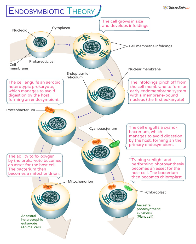
Endosymbiotic theory is the unified and widely accepted theory of how organelles arose in organisms, differing prokaryotic organisms from eukaryotic organisms. In endosymbiotic theory, consistent with general evolutionary theory, all organisms arose from a single common ancestor. This ancestor probably resembled a bacteria, or prokaryote with a single strand of DNA surrounded by a plasma membrane. Throughout time, these bacteria diverged in form and function. Some bacteria acquired the ability to process energy from the environment in novel ways. Photosynthetic bacteria developed the pathways that enabled the production of sugar from sunlight. Other organisms developed novel ways to use this sugar is oxidative phosphorylation , which produced ATP from the breakdown of sugar with oxygen. ATP can then be used to supply energy to other reactions in the cell.

Both of these novel pathways led to organisms that could reproduce at a higher rate than standard bacteria. Other species, not being able to photosynthesis sugars or break them down through oxidative phosphorylation, decreased in abundance until they developed a novel adaptation of their own. The ability of endocytosis , or to capture other cells through the enfolding of the plasma membrane, is thought to have evolved around this time. These cells now had the ability to phagocytize , or eat, other cells. In some cells, the bacteria that were ingested were not eaten, but utilized. By providing the bacteria with the right conditions, the cells could benefit from their excessive production of sugar and ATP. One cell living inside of another is called endosymbiosis if both organisms benefit, hence the name of the theory. Endosymbiotic theory continues further, stating that genes can be transferred between the host and the symbiont throughout time.

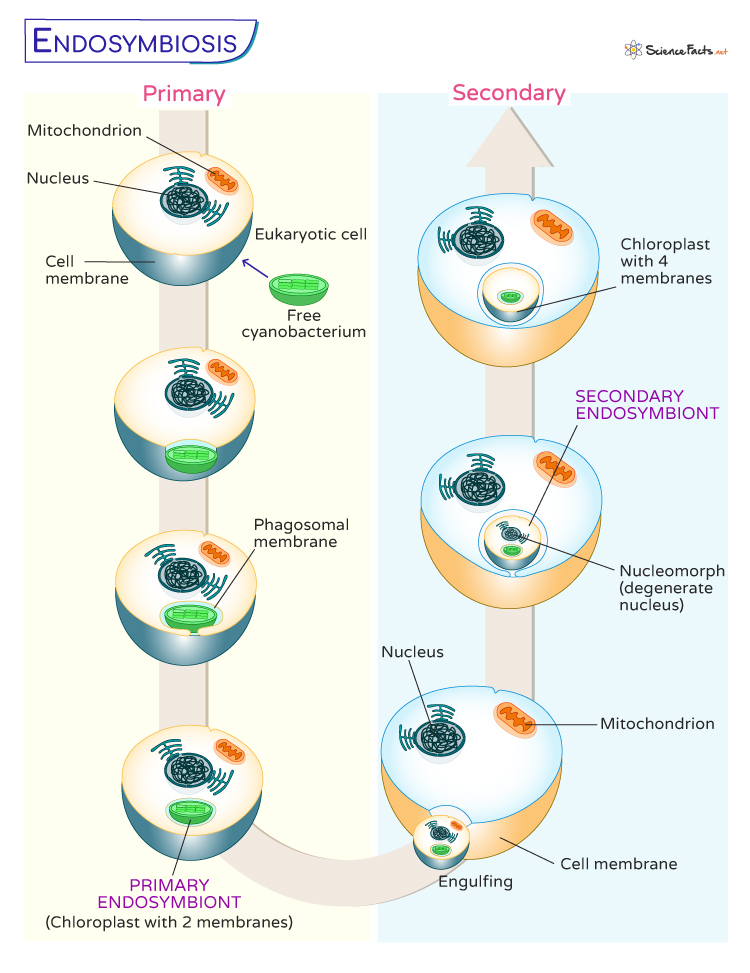
This gives rise to the final part of endosymbiotic theory, which explains the variable DNA and double membranes found in various organelles in eukaryotes. While the majority of cell products start in the nucleus, the mitochondria and chloroplast make many of their own genetic products. The nucleus, chloroplasts, and mitochondria of cells all contain DNA of different types and are also surrounded by double membranes, while other organelles are surrounded by only one membrane. Endosymbiotic theory postulates that these membranes are the residual membranes from the ancestral bacterial endosymbiont. If a bacteria was engulfed via endocytosis, it would be surrounded by two membranes. The theory states that these membranes survived evolutionary time because each organism retained the maintenance of its membrane, even while losing other genes entirely or transferring them to the nucleus. Endosymbiotic theory is supported by a large body of evidence. The general process can be seen in the following graphic.

Serial endosymbiosis

Endosymbiotic Theory Evidence

The most convincing evidence supporting endosymbiotic theory has been obtained relatively recently, with the invention of DNA sequencing. DNA sequencing allows us to directly compare two molecules of DNA, and look at their exact sequences of amino acids. Logically, if two organism share a sequence of DNA exactly, it is more likely that the sequence was inherited through common descent than the sequence arose independently. If two unrelated organisms need to complete the same function, the enzyme they evolve does not have to look the same or be from the same DNA to fill the same role. Thus, it is much more likely that organisms who share sequences of DNA inherited them from an ancestor who found them useful.

This can be seen when analyzing the mitochondrial DNA (mtDNA) and chloroplast DNA of different organisms. When compared to known bacteria, the mtDNA from a wide variety of organisms contains a number of sequences also found in Rickettsiaceae bacteria. Fitting with endosymbiotic theory, these bacteria are obligate intracellular parasites. This means they must live within a vesicle of an organism that engulfs them through endocytosis. Like bacterial DNA, mtDNA and the DNA in chloroplasts is circular. Eukaryotic DNA is typically linear. The only genes missing from the mtDNA and those of the bacteria are for nucleotide, lipid, and amino acid biosynthesis. An endosymbiotic organism would lose these functions over time, because they are provided for by the host cell.

Further analysis of the proteins, RNA and DNA left in organelles reveals that some of it is too hydrophobic to cross the external membrane of the organelle, meaning the gene could never get transferred to the nucleus, as the cell would have no way of importing certain hydrophobic proteins into the organelle. In fact, chloroplasts and mitochondria have their own genetic code, and their own ribosomes to produce proteins. These proteins are not exported from the mitochondria or chloroplasts, but are needed for their functions. The ribosomes of mitochondria and chloroplasts also resemble the smaller ribosomes of bacteria, and not the large eukaryotic ribosomes. This is more evidence that the DNA originated inside of the organelles, and is separate completely from the eukaryotic DNA. This is consistent with endosymbiotic theory.

Lastly, the position and structure of these organelles lends to the endosymbiotic theory. The mitochondria, chloroplasts, and nuclei of cells are all surrounded in double membranes. All three contain their DNA in the center of the cytoplasm, much like bacterial cells. Although less evidence exists linking the nucleus to any kind of extant species, both chloroplasts and mitochondria greatly resemble several species of intracellular bacteria, existing in much the same manner. The nucleus is thought to have arisen through enfolding of the cell membrane, as seen in the graphic above. Throughout the world, there are various endosymbiont bacteria, all of which live inside other organisms. Bacteria exist almost everywhere, from the soil to inside our gut. Many have found unique niches within the cells of other organisms, and this is the basis of endosymbiotic theory.

Related Biology Terms

  • Endosymbiont – An organism that lives with another organism, cause both organisms to receive benefits.
  • Cyanobacteria – Still extant, cyanobacteria are photosynthetic bacteria whose ancestors probably became the chloroplasts of plant cells.
  • Proteobacteria – The bacterial ancestor to the mitochondria organelle.
  • Eukaryote – An organism with membrane bound organelles, thought to have evolved from endosymbiotic interactions.

Cite This Article

Subscribe to our newsletter, privacy policy, terms of service, scholarship, latest posts, white blood cell, t cell immunity, satellite cells, embryonic stem cells, popular topics, pituitary gland, horticulture, hermaphrodite.

What Is the Hygiene Hypothesis?

Viruses and autoimmune disorders

Many parents believe that their children must be kept in an environment that is as clean as possible, but some research suggests that being exposed to what many would call unclean conditions is good for a child's immune system. Research has indicated that children who are kept in very clean environments have a higher rate of hay fever, asthma and a wide range of other conditions. This is what is called the hygiene hypothesis.

The hygiene hypothesis was first introduced in the late 1980s by David P. Strachan, a professor of epidemiology, in the British Medical Journal. Strachan found that children in larger households had fewer instances of hay fever because they are exposed to germs by older siblings. This finding led to further research that suggests a lack of early childhood exposure to less than pristine conditions can increase the individual's susceptibility to disease.

For example, in the late 1990s, Dr. Erika von Mutius, a health researcher, compared the rates of allergies and asthma in East Germany and West Germany, which unified in 1999. Her initial hypothesis was that East German children, who grew up in dirtier and generally less healthful conditions, would have more allergies and suffer more from asthma than their Western counterparts. However, her research found the opposite: children in the polluted areas of East Germany had lower allergic reactions and fewer cases of asthma than children in West Germany. 

Further research has found that children in developing areas of the world are less likely to develop allergies and asthma compared with children in the developed world. 

Building the immune system

The idea is simple. When babies are inside the womb they have a very weak immune system because they are given protection by their mother's antibodies. When they exit the womb, though, the immune system must start working for itself. For the immune system to work properly, it is thought that the child must be exposed to germs so that it has a chance to strengthen, according to the U.S. Food and Drug Administration (FDA). 

The idea is similar to the training of a body builder. For a body builder to be able to lift heavy objects, the muscles must be trained by lifting heavier and heavier objects. If the body builder never trains, then he will be unable to lift a heavy object when asked. The same is thought to be true for the immune system. In able to fight off infection, the immune system must train by fighting off contaminants found in everyday life. Systems that aren't exposed to contaminants have trouble with the heavy lifting of fighting off infections.

Mutius hypothesized that the reason children who are not exposed to germs and bacteria are sicklier is due to how the human immune system evolved. She thinks there are two types of biological defenses. If one of the defense systems isn't trained or practiced enough to fight off illness, the other system overcompensates and creates an allergic reaction to harmless substances like pollen.

Research by other scientists has found similar results. Exposure to germs triggered an internal inflammatory response in children who were raised in cleaner environments, leading to ailments such as asthma, according to a 2002 article in Science magazine.

One researcher has personal experience has leads him to back the hygiene hypothesis. "I believe that there is a role in the development of a child's immunity exposure to various germs and a vast microbiome diversity," said Dr. Niket Sonpal, an assistant professor of clinical medicine at Touro College of Osteopathic Medicine, Harlem Campus. "I was born in India but moved to the U.S. and went to college in Virginia and medical school in Europe. I am sure that the vast change in environment has played a role in my immunity. How has it? I don't think we know just yet." 

In 1997, some began to question if there is a correlation between the hygiene hypothesis and vaccinations. The number of children getting vaccinations was going up, but so were the number of children afflicted with allergies, eczema and other problems. Could depriving the developing immune system of infections using vaccines cause the immune system to eventually attack itself and cause autoimmune diseases like asthma and diabetes? This is a highly contested issue. 

Three studies conducted in the 1990s showed that vaccines had no correlation with children developing allergies and other ailments later in life. In fact, vaccinations may help prevent asthma and other health problems other than the diseases they were intended to prevent, according to The National Center for Immunization Research and Surveillance . The idea that vaccinations can cause health problems does not consider the fact that children, whether vaccinated or not, are still exposed to pathogens that help build the immune system. These pathogens also have no relation to the diseases that the vaccines prevent. 

The conflict between cleanliness and exposure can leave parents feeling confused. There are many microbes that can make children very sick, such as such as respiratory syncytial virus (RSV), E.coli and salmonella. So cleaning the home is still very important. What should children be exposed to and what should they be protected from? 

The CDC recommends regularly cleaning and disinfecting surfaces in the home, especially when surfaces have been contaminated by fecal matter or meat or have come in contact with those who have a virus. Children are also encouraged, though, to play outside , even if they may get dirty in the process. This balancing act may prove to help children stay healthy while still developing a healthy immune system. 

Sonpal thinks that the healthy growth of the immune system isn't just about coming in contact with dirt. It also has to do with what foods are consumed, what kind of environments the person grows up in and intrinsic genetics coupled with physical activity levels. Harvard Medical School noted that getting plenty of sleep, avoiding cigarette smoke, drinking in moderation and controlling blood pressure also all play a part in a healthy immune system.

Additional Resources

  • Clinical & Experimental Immunology: The 'Hygiene Hypothesis' for Autoimmune and Allergic Diseases: An Update
  • Mayo Clinic: Early germ exposure prevents asthma?
  • U.S. National Library of Medicine: The Hygiene Hypothesis and home hygiene

Sign up for the Live Science daily newsletter now

Get the world’s most fascinating discoveries delivered straight to your inbox.

Genetically engineered 'mind control' parasite could deliver drugs to the brain

Eating too much sugar may accelerate cellular aging

'Simone is a very, very rare bird': Experts discuss the science behind Simone Biles' gymnastic prowess

Most Popular

  • 2 What defines a species? Inside the fierce debate that's rocking biology to its core
  • 3 Newly discovered asteroid larger than the Great Pyramid of Giza will zoom between Earth and the moon on Saturday
  • 4 China opens Chang'e 6 return capsule containing samples from moon's far side
  • 5 Neanderthals cared for 6-year-old with Down syndrome, fossil find reveals
  • 2 This robot could leap higher than the Statue of Liberty — if we ever build it properly
  • 3 Zany polar bears and a '3-headed' giraffe star in Nikon Comedy Wildlife Awards
  • 4 Which continent has the most animal species?
  • 5 Newly discovered asteroid larger than the Great Pyramid of Giza will zoom between Earth and the moon on Saturday

what is a hypothesis for bacteria

April 7, 2011

Can It Be Bad to Be Too Clean?: The Hygiene Hypothesis

Johns Hopkins School of Medicine researcher Kathleen Barnes talks about the hygiene hypothesis, which raises the possibility that our modern sterile environment may contribute to conditions such as asthma and eczema

By Steve Mirsky

what is a hypothesis for bacteria

Johns Hopkins School of Medicine researcher Kathleen Barnes talks about the hygiene hypothesis , which raises the possibility that our modern sterile environment may contribute to conditions such as asthma and eczema.

Podcast Transcription

Steve:          Welcome to Science Talk , the more of less weekly podcast of Scientific American , posted on April 6th, 2011. I am Steve Mirsky. This week on the podcast:

On supporting science journalism

If you're enjoying this article, consider supporting our award-winning journalism by subscribing . By purchasing a subscription you are helping to ensure the future of impactful stories about the discoveries and ideas shaping our world today.

Barnes:          The hypothesis is that as we make the shift from dirt to sterile that you're changing the direction of your immune response. This causes diseases.

Steve:          That's Kathleen Barnes. We'll hear from her, and we'll test your knowledge of some recent science in the news. Kathleen Barnes studies what's called the hygiene hypothesis at the Johns Hopkins School of Medicine in Baltimore. She presented some of her research at the recent meeting of the American Association for the Advancement of Science in Washington, D.C., after which we sat down to chat.

Steve:          First, tell me what is the hygiene hypothesis, for people who haven't heard of it?

Barnes:          So, the hygiene hypothesis, I guess, simply stated is this notion that as human society has morphed from developing environment, or what we consider a developing world environment, into the developed world, there have been radical changes in our environment; changes associated with the size of families, so going from many to fewer siblings. The idea being that with fewer children in the house, there's less opportunity for exposure to viruses. The idea that we move from a rural to an urban environment; that we have moved from that situation where we're exposed to microbes—one of the best examples in asthma being the idea that we're exposed to endotoxins that is a byproduct of the livestock and farms—to moving to an environment that's more sterile, where we don't have such exposures. The notion that as we move from a developing to a developed environment, we have less exposure to microbes in general. We treat every symptom with antibiotics; we've changed our gut microflora with the diets that we eat.

Steve:          We use antibacterial soap and antibacterial surface cleaners in the house.

Barnes:          Exactly, yeah, the idea is sterile is good. Sterile is healthy.

Steve:          That's the hygiene; what's the hypothesis part?

Barnes:          The hypothesis is that as we make the shift from dirt to sterile that you are changing the direction of your immune response. And so in the context of asthma, and frankly in other autoimmune diseases and diseases of inflammation, it's this imbalance from that side of our immune response that we believe evolved to protect us against things like bacteria and viruses and malarial parasites to the other side of our immune system that, frankly, when it's revved up causes diseases like allergies and some of these other diseases of inflammation. So it's really this imbalance between these two sides of our immune system, both which were designed to do something good for us; but when it's not equal, when it's imbalanced, we're going to have too much of one disease versus another.

Steve:          So without the exposure to these environmental challenges, we wind up trading one set of conditions for a different set of conditions or illnesses.

Barnes:          Exactly. And the whole, the notion of the epidemiological transition is that we've gone from a situation in our distant past, where we're exposed to lots of microbes—but we also, to be frank, we died from a lot of these diseases; so, it's not to romanticize our past, certainly these microbes were able to kill us and cause great consternation in city-state populations. But the idea is that some balance protects you on the one hand from some of those.

Steve:          Yeah, I mean, nobody wants to go back to the days when we didn't have clean drinking water. Arguably clean drinking water is the single most important public health development in the history of humanity. And so we're not dying so much in the developed world of dysentery and other conditions of unsanitary drinking water, and we don't have to drink alcohol all day to make sure that we're not drinking dangerous liquids; but you know, dangerous in a different way and people used to die younger because they were getting infection and you would die from it. And, I mean, that still those happen today but, you know, fortunately we do have antibiotics and other things. So we're not arguing to go back to that. But we have realized that people have teased out the fact that maybe some of the now epidemic asthma rates, for example, are related to the fact that kids when they are growing up and even before they're born are not being challenged, their immune systems are not being challenged the way that we evolved.

Barnes:          So I was going to throw that very example out, sort of, the classic example or the classic notion is that an individual's propensity to be more upregulated on one side of their immune system—say Th1 versus their Th2, the Th1 side being that side of our immune system that we believe evolved to protect us against things like the bacteria and the viruses and microbes; versus Th2 which served a very fundamental purpose in an environment where one was exposed to worms. But we're not exposed to worms anymore, so that side doesn't serve as much of a purpose, and we know from very sophisticated studies that infants that are born to mothers in the, sort of, with the, sort of, sterile environment, infants are born with the predisposition to be Th2 skewed. And there is a reason for that. During the neonatal period, it's important for the mother not to reject the fetus as the fetus is developing and so the mother's immune response is slightly tilted towards this Th2 to not treat the fetus as a microbe, if I can put it that bluntly.

Steve:          Which is really a good thing.

Barnes:          (laughter) At the time that when the infant is born, so the infant is born with a slight Th2 preference over the Th1, because that was the intrauterine environment. The thinking is that when exposed to some bacteria, some viruses, it sort of shifts this back into an equilibrium. But unfortunately in our current environment, where everything is sterile, we tend to forego breastfeeding and feed our infants formula from sterile water and so on and so forth, there are fewer siblings at home, we're not really giving these infants the chance to equalize, if you will, that immune response.

Steve:          So what are some of the actual studies? What are your research interests that have confirmed that this situation exists out there?

Barnes:          So, there have been a number of studies in the field of asthma, studies that have not necessarily been our own certainly include the German farming studies by Erika von Mutius and others showing that children who lived in very close proximity to livestock, and we know are exposed to lots of bacteria, for example, from the livestock are less likely to have asthma and allergies. There have been beautiful studies showing that children who go to daycare very early in life, who are therefore exposed to more rhinovirus and virus in general tend to have fewer asthma and allergies as they grow up. In our own work, we've taken a closer look at that exposure to that bacteria, the gram-negative bacteria we call endotoxin, which is ubiquitous, it's around us all the time; but it's certainly higher in some places than others and endotoxin we know doesn't just come from livestock, endotoxin comes from diesel exhaust. So there have been some various nice studies showing elevated levels of endotoxins in regions where folks live in close proximity to traffic. Right up the road in the Baltimore tunnel, there's some of the highest endotoxin levels that have ever been recorded.

Steve:          How does that it get in the diesel exhaust?

Barnes:          It's part of the particulate matter.

Steve:          It's just picking it up from the environment and pluming it out?

Barnes:          Exactly, we were interested in testing that theory that with higher levels of endotoxin, there would be lower levels of asthma and allergy. When we measure endotoxin in the tropical environment it looks very different than it does in a developed environment such as here. It's much higher and it's probably higher for a variety of reasons. In our one particular study that we've done in Barbados, folks live very close to the road, and they're exposed to very high levels of diesel exhaust, and we believe that pollution has contributed over time to an ever increasing prevalence of asthma in that society. When we measure endotoxin in the homes of these folks, it's very, very high but they have a lot of asthma and allergy, so I think that's telling us that the hygiene hypothesis is not a black and white hypothesis, that there are a lot of complexities to this. And for me personally it tells me that there are also genetic underpinnings. So it's not just about exposure to the environment, just as it's not just about having a particular mutation that puts you at risk of developing disease, but it's the interaction of those two factors—genes and environment that will probably help us have a better understanding of the Hygiene hypothesis.

Steve:          It's definitely multifactorial, but when you deal with large enough populations you can start to tease out these kinds of relationships.

Barnes:          Absolutely. Having the opportunity to study very large populations allows you to stratify folks based on exposure to factor A and B; allows you to stratify on not just one genetic polymorphism but many polymorphisms. So there is much to be gained from these very large population studies. And I frankly think that to get a better handle on the role of the hygiene hypothesis not just an asthma and allergic disease but any of these other complex diseases, the real key also are longitudinal studies, birth cohort studies, where we've been able to track an individual from the time he or she is born, measure various environmental exposures along the way, and then compare that to their genetic background.

Steve:          So, we're not going to advise people to, oh go get the helminth worm infection, or you know, go roll around in the dirt on a farm nearby with your kids; nobody is going to advise that. So what is the practical application going to be of this kind of knowledge?

Barnes:          Right, so it's very tempting, it's very tempting to come up with a conclusion that being exposed to a lot of dirt is good for your health or that being exposed to and infested with worms is good for your health. We wouldn't advocate either of those, but what I think the real value in this science really is, is if we can understand what it is about those microbes, and in the case of the parasite, what is it about the protein within the parasite that elicits this response to the parasite, either protective or conferring risk of the disease? If we could put our finger on that particular molecule we could develop better therapeutics for people. We could, you're not going to advocate giving an individual worms to cure their asthma or to cure their autoimmune disease. But you could come up with a drug that has some part of that protein that elicits this biological response and give that to the individual therapeutically.

Steve:          How did you wind up working on this subject? Did you start way back in grad school? Or I don't mean way back.

Barnes:          (laughter) No, you can say that because it's true.

Steve:          Okay, or was it something that came along in the middle of your academic career?

Barnes:          My work has really evolved truly over several decades. So as a graduate student, I was very interested, as a graduate student in medical anthropology, in differences across human populations and our response to environmental factors that confer risk of disease; and schistosomiasis was one disease I was particularly interested in.

Steve:          This is a worm-borne disease.

Barnes:          Schistosomiasis is a worm-borne disease; it's referred to as a helminth and it's spread to the human host through infested waters. Schistosomiasis typically doesn't carry a very high mortality rate. It can be debilitating for those individuals who can't mount an appropriate response against the worm. But my interest really started at that point in time with this interest in how and why we respond to different factors such as parasites, and truly that dovetailed in to what was a growing interest in the mid '80s of why there was an increase in asthma and allergic disease; and the hypothesis had been put forth that the IgE antibody that we all make but typically in very low quantities, had only been discovered in 1968. So, it was a relatively new immunological molecule that we knew was important in our immune system, and we knew it was very important in causing risk to asthma and other allergies like eczema and hay fever. At the same time, we began to appreciate that this IgE molecule was also protective against extracellular parasites. So, I was interested in that co-association with this molecule IgE. Over time, my research really focused more on identifying genetic determinants for asthma and allergies. I put the schistosomiasis studies aside. And then about 10 years ago, I had the opportunity to join forces with colleagues and immunologists in Brazil where schistosomiasis is still quite endemic and; in fact it's one of the last strongholds for schistosomiasis in the western hemisphere. And it just provided a really unique opportunity to test the hypothesis that some of these asthma genes that we had identified might also be important in schistosomiasis.

Steve:          This is, you know, the hackneyed question, but where do you think things might be in another 10 or 15 years?

Barnes:          I think that the hygiene hypothesis particularly and a sort of Darwinian approach in the way we think about medicine in general; that is how did we develop heart disease? How did we develop allergic diseases? How did we develop inflammatory bowel disease? We know as anthropologists that traditional foraging societies didn't have these diseases, and we know that it is not just because they didn't live past the age of 40. They simply didn't develop many of these complex chronic diseases that we experience now. So, I think this new way of approaching the pathology behind these different diseases is opening our minds and understanding, or elucidating new pathways that we didn't think about before. And I am very hopeful that with the better understanding of the pathologies that contribute to these diseases, with a better understanding of how a genetic mutation long ago that protected us against one disease now coincidentally causes another disease, will absolutely help us to develop better therapeutic targets; and not just treat these diseases but also predict who is going to be at greatest risk for disease. And once we know enough about all the environmental factors that go into causing a disease, we as medical folk can give better advise to patients about what they need to avoid or how they need to modify their lifestyle so that they can live longer and healthier lives.

Steve:          This host-pathogen interaction is really so fascinating to study because you're dealing with this co-evolutionary situation but the rates are so different. So, we're practically standing still compared to the rates at which the pathogens get to evolve. And it's just I think, it's one of the most fascinating fields to be studying right now.

Barnes:          It's indeed an exciting field, and I think it's a very important perspective that where we are now in the 21st century with the diseases we face, is such a recent moment in our human evolutionary time. And frankly that's one of the reasons why we've been particularly interested in focusing on diseases for which there is tremendous ethnic and racial disparities. Because it really isn't until very recently in time that we see the admixture, the mixing of individuals of different ancestral backgrounds. And it actually provides us a great opportunity to try to tease out at least from a genetic epidemiology perspective what mutations might have evolved that were selective or advantageous in one particular environment that individuals have carried with them as they've migrated to new parts of the world; and basically admix with other populations for which there might not have been selective advantage for having a mutation and therefore no mutation at all. And so by studying populations from a more ancient background, if I could, we believe that because our gene pool simply hasn't had enough time to change this rapidly, it's a great test tube experiment, if you will.

Steve:          Natural experiment….

Barnes:          A natural experiment to say, okay just yesterday in the time clock of human evolution you lived in an environment where if you have this mutation you will better off than the next guy who didn't have it, because it protected you against, for example, parasites. But now you've moved to another part of the world, and you're not exposed to those parasites anymore, but you still have that mutation that was adaptive back in that environment. Since you're not there anymore and you're exposed to new environmental factors, does that place you at risk for diseases that your ancestors wouldn't have thought about?

Steve:          It's really fascinating. And we may be living in a unique window of time to do those studies, because with the mixing of all the different human population groups, in another couple of hundred years, it may be impossible to do those kinds of studies.

Barnes:          So this is a really important point that right now it's a really terrific opportunity for us to try to tease out what about our human genome, based on our bio-geographical past, can we look at to try to explain why in contemporary times we have disease X. But this will change over time because with that mixture, there are simply diminishing of the benefit of having these mutations that might have been adaptive in a previous time. So yes it is a unique opportunity. And I guess I will just add to that the other opportunity, if we can call it that, is that with globalization and rapid changes in these places that we perform these studies where we are looking at more traditional ways of living and then comparing that to the way we live here in the United States, for example; so even within our Brazil study where we've studied parasitic disease, certainly the public health goal is to eradicate parasitic disease in these populations. And that is the first and foremost priority. But as that happens, we will have less and less opportunity to understand what it is about our past that brought us to where we are now in terms of these genetic polymorphisms that might have served some beneficial purpose.

Steve:          Right. We're in no way saying not to try to make this situation better. We're just saying that we better do these studies now before we do accomplish what we hope to accomplish.

Barnes:          Exactly. We can learn so much from our past by comparing people who live in different environments under different conditions and in different degrees of development, and I think that comes back to the hygiene hypothesis—that it is a unique opportunity to compare populations with different degrees of development to try to hone in on those specific factors that were emblematic of our past but are simply not part of our modern lifestyle.

Steve:          Now it's time to play TOTALL……. Y BOGUS. Here are four science stories; only three are true. See if you know which story is TOTALL……. Y BOGUS.

Story 1: In the newest version of the popular Madden NFL football video game players can get concussions.

Story 2: The late Elizabeth Taylor probably had a FOXC2 gene mutation that affects embryonic development.          Story 3: Wind turbines are becoming a huge killer of birds, taking down some 20 million annually.

And story 4: Growing salamander embryos have been found to have algae living happily inside their tissues. While you think about those stories: Do you believe in the effectiveness of subliminal messages? I think the idea that such messages can really work has pretty much been discredited, but who knows.

Hey, you're time is up.

Story 1 is true. In the new Madden Football game, players can get concussed and will be unavailable for the remainder of the game. So as in real football the object of the game will be to give the opposing team's quarterback a concussion.

Story 2 is true. Liz Taylor probably did have the FOXC2 gene mutation which conferred upon her a double role of her famous thick eyelashes.

And story 4 is true. Growing salamander embryos have been found to harbor algae within their tissues. Researchers think the algae get nitrogen rich waste products and the salamanders get oxygen. It's a winner-winner salamander dinner situation. For more check out the April 5th episode of the daily SciAm, podcast, 60-Second Science .

All of which means that Story 3 about wind turbines killing 20 million birds annually is TOTALL……. Y BOGUS. Now what is true is that the bird death toll from turbines is significant—about 440,000 birds per year according to the US Fish and Wildlife Service, and it's an issue that needs to be addressed as wind power hopefully becomes more of an energy contributor. But the real bird killer is on your couch. The American Bird Conservancy says that domestic cats kill about 250 million birds each year, a figure matched by feral cats; which means that wind turbines right now get less than a 10th of a percent as many birds as cats do.

That's it for this episode. Get your science news at www.ScientificAmerican.com, where you can check out the Daily Science Agenda, which features what you need to know now. For example: How does a 737 lose its fuselage in mid-flight? Find out at our Web site safely on the ground and follow us on Twitter, where you'll get a tweet about each new article posted to our Web site. Our Twitter handle is @sciam. For Science Talk , the podcast of Scientific American , I'm Steve Mirsky. Thanks for clicking on us. Try the Scientific American smart phone app.

what is a hypothesis for bacteria

What is the Microbial Diversity Hypothesis?

  • Download PDF Copy

Yolanda Smith, B.Pharm.

The microbial diversity hypothesis suggests that the diversity and turnover of bacterial species in the gut mucosa and other areas around the body are key factors in the regulation of the immune system. This is in contrast to the historical belief that the body showed stable colonization with certain microbial species.

It was proposed as an extension of or alternative to the hygiene hypothesis, following the emergence of several factors that could not be explained by the original hypothesis.

Shortcomings of the Hygiene Hypothesis

The hygiene hypothesis, as initially proposed by Strachan in 1989, traced the supposed reduction in exposure to microbes during childhood to the rise in the incidence of allergic and autoimmune disease .

However, this theory is unable to explain several factors about the epidemiology of allergic disease including:

  • Why allergic asthma is on the rise in American cities classed as 'unhygienic'.
  • Why migrant children in some large European cities have a lower incidence of allergic disease despite sharing many common characteristics as to the environment.
  • Why infection by airborne viruses does not seem to protect from allergic sensitization.
  • Why research has not shown congruent findings to support the link between certain viral infections and allergic diseases in diverse populations.
  • The inefficiency of probiotics in the prevention and treatment of allergic diseases.

As a result of these factors, there is a need to reconsider the basis of the hygiene hypothesis and identify particular microbial agents that may play a protective role in the prevention of allergic diseases.

Microbial Diversity

The microbial diversity, also known as the “high turnover and diversity hypothesis”, was first proposed by Paolo Matricardi, and later refined by von Herzen.

It suggests that a high turnover of bacteria at the mucosal level, located in the lymphoid tissue of the nasal tubes, bronchi and gut, is the predominant state of the normal body. This is in contrast to the supposed static colonization by particular species. This wide and changing variety of bacteria causes the presentation of many strong antigens to the immune system, resulting in its incessant stimulation. This is thought to have a protective effect against atopic and allergic diseases.

Some research has supported this notion, although the sample size was small and further studies are required to validate the hypothesis. It remains unclear if the protective effect is brought about by diversity alone, or by a diverse population which encompasses particular organisms that are necessary for the development of the immune system.

Related Stories

  • Experts publish guidance to help safely and equitably implement Palforzia peanut oral immunotherapy
  • Innovative HIV vaccine approaches yield potential for broad protection against viral strains
  • Long-term immune changes persist in unvaccinated COVID-19 patients

The embryonic immune system has been compared to a computer that is equipped with programs but lacks significant data. As individuals progress through gestation and infancy, they are naturally exposed to a number of diverse organisms to fill up their database of organisms that can be recognized by the immune system. This allows them to identify known and harmful agents, and to exhibit an allergic response as deemed necessary.

Allergic Disease

The microbial diversity hypothesis is of particular interest in that it seems likely that a lack of diversity may be linked to an increased incidence of allergic disease. Specific life stages are considered to be the most important points in regards to the development of allergies. These include:

  • Early development.
  • Later part of pregnancy.
  • The first days and months of infancy.

For this reason, it is essential that microbial exposure is maintained over an extended period of time that covers these critical life stages.

In fact, there is some research to suggest that being born vaginally has a protective effect against the development of allergic disease. It is postulated that this may be linked to the passage of natural flora that are passed from mother to infant during birthing. In contrast, infants delivered by Caesarean section are more likely to be affected by allergies. However, breastfeeding has long been proved to help reduce the risk of allergies for all babies, regardless of the mode of delivery.

Beyond birth and through the early years of childhood, the effect of microbial exposure required for optimal outcomes remains unknown.

  • http://ifh-homehygiene.org/
  • http://onlinelibrary.wiley.com/doi/10.1111/1462-2920.12895/abstract
  • http://www.ncbi.nlm.nih.gov/pmc/articles/PMC2841842/      

Further Reading

  • All Allergy Content
  • What are Allergies?
  • Different Types of Allergies
  • Old Friends Hypothesis
  • Allergies and the Hygiene Hypothesis

Last Updated: Dec 30, 2022

Yolanda Smith

Yolanda Smith

Yolanda graduated with a Bachelor of Pharmacy at the University of South Australia and has experience working in both Australia and Italy. She is passionate about how medicine, diet and lifestyle affect our health and enjoys helping people understand this. In her spare time she loves to explore the world and learn about new cultures and languages.

Please use one of the following formats to cite this article in your essay, paper or report:

Smith, Yolanda. (2022, December 30). What is the Microbial Diversity Hypothesis?. News-Medical. Retrieved on August 01, 2024 from https://www.news-medical.net/health/What-is-the-Microbial-Diversity-Hypothesis.aspx.

Smith, Yolanda. "What is the Microbial Diversity Hypothesis?". News-Medical . 01 August 2024. <https://www.news-medical.net/health/What-is-the-Microbial-Diversity-Hypothesis.aspx>.

Smith, Yolanda. "What is the Microbial Diversity Hypothesis?". News-Medical. https://www.news-medical.net/health/What-is-the-Microbial-Diversity-Hypothesis.aspx. (accessed August 01, 2024).

Smith, Yolanda. 2022. What is the Microbial Diversity Hypothesis? . News-Medical, viewed 01 August 2024, https://www.news-medical.net/health/What-is-the-Microbial-Diversity-Hypothesis.aspx.

Suggested Reading

Health experts call for a renewed focus on allergy training for nurses

Cancel reply to comment

  • Trending Stories
  • Latest Interviews
  • Top Health Articles

Can cutting back on extra virgin olive oil make your heart healthier?

A Discussion with Hologic’s Tim Simpson on the Future of Cervical Cancer Screening

Tim Simpson

Hologic’s Tim Simpson Discusses the Future of Cervical Cancer Screening.

A Discussion with Hologic’s Tim Simpson on the Future of Cervical Cancer Screening

From Waste to Taste: The Transformative Power of Fermented Foods

Maria Marco

In this interview conducted at Pittcon 2024 in San Diego, Maria Marco discusses her research on the health benefits, safety, and waste reduction potential of fermented foods, and the microbial processes involved in their production.

From Waste to Taste: The Transformative Power of Fermented Foods

Revolutionizing hepatocyte count for researchers

Dan Schieffer

In this interview, NewsMedical speaks to DeNovix's Dan Schieffer about the new app developed for CellDrop, and how it aims to revolutionize hepatocyte count for the drug discovery industry, and more.

Revolutionizing hepatocyte count for researchers

Latest News

Study reveals obesity's impact on muscle structure in heart failure with preserved ejection fraction

Newsletters you may be interested in

Asthma

Your AI Powered Scientific Assistant

Hi, I'm Azthena, you can trust me to find commercial scientific answers from News-Medical.net.

A few things you need to know before we start. Please read and accept to continue.

  • Use of “Azthena” is subject to the terms and conditions of use as set out by OpenAI .
  • Content provided on any AZoNetwork sites are subject to the site Terms & Conditions and Privacy Policy .
  • Large Language Models can make mistakes. Consider checking important information.

Great. Ask your question.

Azthena may occasionally provide inaccurate responses. Read the full terms .

While we only use edited and approved content for Azthena answers, it may on occasions provide incorrect responses. Please confirm any data provided with the related suppliers or authors. We do not provide medical advice, if you search for medical information you must always consult a medical professional before acting on any information provided.

Your questions, but not your email details will be shared with OpenAI and retained for 30 days in accordance with their privacy principles.

Please do not ask questions that use sensitive or confidential information.

Read the full Terms & Conditions .

Provide Feedback

what is a hypothesis for bacteria

If you're seeing this message, it means we're having trouble loading external resources on our website.

If you're behind a web filter, please make sure that the domains *.kastatic.org and *.kasandbox.org are unblocked.

To log in and use all the features of Khan Academy, please enable JavaScript in your browser.

AP®︎/College Biology

Course: ap®︎/college biology   >   unit 2.

  • Prokaryotic and eukaryotic cells
  • Intro to eukaryotic cells
  • Endomembrane system
  • The endomembrane system

Endosymbiosis theory

  • Mitochondria and chloroplasts
  • Prokaryotes and eukaryotes review
  • Cell compartmentalization

what is a hypothesis for bacteria

Want to join the conversation?

  • Upvote Button navigates to signup page
  • Downvote Button navigates to signup page
  • Flag Button navigates to signup page

Good Answer

Video transcript

More From Forbes

What is ‘panspermia’ new evidence for the wild theory that says we could all be space aliens.

  • Share to Facebook
  • Share to Twitter
  • Share to Linkedin

"Lithopanspermia" involves a comet or asteroid striking a planet, in this case Mars, which then ... [+] ejects biologically-rich matter into space, eventually settling on Earth. (Illustration by Tobias Roetsch/Future Publishing via Getty Images)

Imagine if NASA’s Mars Perseverance rover—now on its way to the red planet— discovered microbial life there.

It would change everything we know about life in the Solar System and far beyond.

Or would it? What if we accidentally transported life to Mars on a spacecraft? And what if that is how life moves around the Universe?

A new paper published this week in Frontiers in Microbiology explores the possibility that microbes and extremophiles may migrate between planets and distribute life around the Universe—and that includes on spacecraft sent from Earth to Mars.

This is the controversial theory of “panspermia.”

Best Travel Insurance Companies

Best covid-19 travel insurance plans, what is ‘panspermia’.

It’s an untested, unproven and rather wild theory regarding the interplanetary transfer of life. It theorizes that microscopic life-forms, such as bacteria, can be transported through space and land on another planet. Thus sparking life elsewhere.

It could happen by accident—such as on spacecraft—via comets and asteroids in the Solar System, and perhaps even between star systems on interstellar objects like ʻOumuamua .

However, for “panspermia” to have any credence requires proof that bacteria could survive a long journey through the vacuum, temperature fluctuations, and intense UV radiation in outer space.

Cue the “Tanpopo” project.

The bacterial exposure experiment took place from 2015 to 2018 using the Exposed Facility located on ... [+] the exterior of Kibo, the Japanese Experimental Module of the International Space Station.

What is the ‘Tanpopo’ mission?

Tanpopo— dandelion in English—is a scientific experiment to see if bacteria can survive in the extremes of outer space.

The researchers from Tokyo University—in conjunction with Japanese national space agency JAXA—wanted to see if the bacteria deinococcus could survive in space, so had it placed in exposure panels on the outside of the International Space Station (ISS). It’s known as being resistant to radiation. Dried samples of different thicknesses were exposed to space environment for one, two, or three years and then tested to see if any survived.

They did, largely by a layer of dead bacteria protecting a colony beneath it. The researchers estimate that a colony of 1 mm of diameter could potentially survive up to 8 years in outer space conditions.

What does this mean for ‘panspermia?’

“The results suggest that deinococcus could survive during the travel from Earth to Mars and vice versa, which is several months or years in the shortest orbit,” said Akihiko Yamagishi, a Professor at Tokyo University of Pharmacy and Life Sciences and principal investigator of Tanpopo.

That means spacecraft visiting Mars could theoretically carry microorganisms and potentially contaminate its surface.

However, this isn’t just about Earth and Mars—the ramifications of panspermia, if proven, are far-reaching.

“The origin of life on Earth is the biggest mystery of human beings (and) scientists can have totally different points of view on the matter,” said Dr. Yamagishi. “Some think that life is very rare and happened only once in the Universe, while others think that life can happen on every suitable planet.”

“If panspermia is possible, life must exist much more often than we previously thought.”

What is ‘lithopanspermia?’

This is bacteria surviving in space for a long period when shielded by rock—typically an asteroid or a comet—which could travel between planets, potentially spreading bacteria and biologically-rich matter around the Solar System.

However, the theory of panspermia goes even further than that.

What is ‘interstellar panspermia’ and ‘galactic panspermia?’

This is the hypothesis—and it’s one with zero evidence—that life exists throughout the galaxy and/or Universe specifically because bacteria and microorganisms are spread around by asteroids, comets, space dust and possibly even interstellar spacecraft from alien civilizations.

In 2018 a paper concluded that the likelihood of Galactic panspermia is strongly dependent upon the survival lifetime of the organisms as well as the velocity of the comet or asteroid—positing that the entire Milky Way could potentially be exchanging biotic components across vast distances.

Such theories have gained credence in the last few years with the discovery of two extrasolar objects Oumuamua and Borisov passing through our Solar System.

However, while the ramifications are mind-boggling, panspermia is definitely not a proven scientific process.

There are still many unanswered questions about how the space-surviving microbes could physically transfer from one celestial body to another.

Illustration of NASA's Mars 2020 Perseverance rover studying a Mars rock outcrop (not to scale). ... [+] Mars 2020 is targeted for launch in July/August 2020 aboard an Atlas V-541 rocket from Space Launch Complex 41 at Cape Canaveral Air Force Station in Florida.

How will Perseverance look for life on Mars?

NASA’s Perseverance rover is due to land on the red planet on February 18, 2021. It will land in a nearly four billion-year-old river delta in Mars’ 28 miles/45 kilometers-wide Jezero Crater. 

It’s thought likely that Jezero Crater was home to a lake as large as Lake Tahoe more than 3.5 billion years ago. Ancient rivers there could have carried organic molecules and possibly even microorganisms.

Perseverance’s mission will be to analyze rock and sediment samples to see if Mars may have had conditions for microorganisms to thrive. It will drill a few centimeters into Mars and take core samples, then put the most promising into containers. It will then leave them on the Martian surface to be later collected by a human mission in the early 2030s. 

Wishing you clear skies and wide eyes.

Jamie Carter

  • Editorial Standards
  • Reprints & Permissions
  • Patient Care & Health Information
  • Diseases & Conditions

Escherichia coli (E. coli) bacteria normally live in the intestines of healthy people and animals. Most types of E. coli are harmless or cause relatively brief diarrhea. But a few strains, such as E. coli O157:H7, can cause severe stomach cramps, bloody diarrhea and vomiting.

You may be exposed to E. coli from contaminated water or food — especially raw vegetables and undercooked ground beef. Healthy adults usually recover from infection with E. coli O157:H7 within a week. Young children and older adults have a greater risk of developing a life-threatening form of kidney failure.

Signs and symptoms of E. coli O157:H7 infection usually begin three or four days after exposure to the bacteria. But you may become ill as soon as one day after exposure to more than a week later. Signs and symptoms include:

  • Diarrhea, which may range from mild and watery to severe and bloody
  • Stomach cramping, pain or tenderness
  • Nausea and vomiting, in some people

When to see a doctor

Contact your doctor if your diarrhea is persistent, severe or bloody.

There is a problem with information submitted for this request. Review/update the information highlighted below and resubmit the form.

From Mayo Clinic to your inbox

Sign up for free and stay up to date on research advancements, health tips, current health topics, and expertise on managing health. Click here for an email preview.

Error Email field is required

Error Include a valid email address

To provide you with the most relevant and helpful information, and understand which information is beneficial, we may combine your email and website usage information with other information we have about you. If you are a Mayo Clinic patient, this could include protected health information. If we combine this information with your protected health information, we will treat all of that information as protected health information and will only use or disclose that information as set forth in our notice of privacy practices. You may opt-out of email communications at any time by clicking on the unsubscribe link in the e-mail.

Thank you for subscribing!

You'll soon start receiving the latest Mayo Clinic health information you requested in your inbox.

Sorry something went wrong with your subscription

Please, try again in a couple of minutes

Only a few strains of E. coli trigger diarrhea. The E. coli O157:H7 strain belongs to a group of E. coli that produces a powerful toxin that damages the lining of the small intestine. This can cause bloody diarrhea. You develop an E. coli infection when you ingest this strain of bacteria.

Unlike many other disease-causing bacteria, E. coli can cause an infection even if you ingest only small amounts. Because of this, you can be sickened by E. coli from eating a slightly undercooked hamburger or from swallowing a mouthful of contaminated pool water.

Potential sources of exposure include contaminated food or water and person-to-person contact.

Contaminated food

The most common way to get an E. coli infection is by eating contaminated food, such as:

  • Ground beef. When cattle are slaughtered and processed, E. coli bacteria in their intestines can get on the meat. Ground beef combines meat from many different animals, increasing the risk of contamination.
  • Unpasteurized milk. E. coli bacteria on a cow's udder or on milking equipment can get into raw milk.
  • Fresh produce. Runoff from cattle farms can contaminate fields where fresh produce is grown. Certain vegetables, such as spinach and lettuce, are particularly vulnerable to this type of contamination.

Contaminated water

Human and animal stool may pollute ground and surface water, including streams, rivers, lakes and water used to irrigate crops. Although public water systems use chlorine, ultraviolet light or ozone to kill E. coli , some E. coli outbreaks have been linked to contaminated municipal water supplies.

Private water wells are a greater cause for concern because many don't have a way to disinfect water. Rural water supplies are the most likely to be contaminated. Some people also have been infected with E. coli after swimming in pools or lakes contaminated with stool.

Personal contact

E. coli bacteria can easily travel from person to person, especially when infected adults and children don't wash their hands properly. Family members of young children with E. coli infection are especially likely to get it themselves. Outbreaks have also occurred among children visiting petting zoos and in animal barns at county fairs.

Risk factors

E. coli can affect anyone who is exposed to the bacteria. But some people are more likely to develop problems than are others. Risk factors include:

  • Age. Young children and older adults are at higher risk of experiencing illness caused by E. coli and more-serious complications from the infection.
  • Weakened immune systems. People who have weakened immune systems — from AIDS or from drugs to treat cancer or prevent the rejection of organ transplants — are more likely to become ill from ingesting E. coli .
  • Eating certain types of food. Riskier foods include undercooked hamburger; unpasteurized milk, apple juice or cider; and soft cheeses made from raw milk.
  • Time of year. Though it's not clear why, the majority of E. coli infections in the U.S. occur from June through September.
  • Decreased stomach acid levels. Stomach acid offers some protection against E. coli . If you take medications to reduce stomach acid, such as esomeprazole (Nexium), pantoprazole (Protonix), lansoprazole (Prevacid) and omeprazole (Prilosec), you may increase your risk of an E. coli infection.

Complications

Most healthy adults recover from E. coli illness within a week. Some people — particularly young children and older adults — may develop a life-threatening form of kidney failure called hemolytic uremic syndrome.

No vaccine or medication can protect you from E. coli -based illness, though researchers are investigating potential vaccines. To reduce your chance of being exposed to E. coli , avoid swallowing water from lakes or pools, wash your hands often, avoid risky foods, and watch out for cross-contamination.

  • Mayo Clinic Minute: Avoiding summer E. coli infection

Cook this incorrectly, and you could end up with a case of E. coli .

" E. coli stands for Escherichia coli, which is a type of bacteria."

"Most commonly, we hear about it in raw or undercooked hamburger meat."

Dr. Nipunie Rajapakse says E. coli bacteria can create some stomach-turning symptoms, like abdominal pain and nausea. But it can get even worse.

"There's a specific type of E. coli . It's called O157:H7, which can cause bloody diarrhea and has been associated with a condition that can cause kidney damage, especially in young children."

The elderly are also at higher risk for problems with E. coli , as are pregnant women, people with underlying digestive problems and those with weakened immune systems.

"If somebody were to be exposed to E. coli in something they ate or drank, they may have symptom onset within a couple of days to a few weeks after infection or exposure."

Dr. Rajapakse says the best way to avoid a bout with the bacteria is to wash your hands and thoroughly cook your hamburgers.

For the Mayo Clinic News Network, I'm Jeff Olsen.

Risky foods

  • Cook hamburgers until they're 160 F (71 C). Hamburgers should be well-done, with no pink showing. But color isn't a good guide to know if the meat is done cooking. Meat — especially if grilled — can brown before it's completely cooked. Use a meat thermometer to ensure that meat is heated to at least 160 F (71 C) at its thickest point.
  • Drink pasteurized milk, juice and cider. Any boxed or bottled juice kept at room temperature is likely to be pasteurized, even if the label doesn't say so. Avoid any unpasteurized dairy products or juice.
  • Wash raw produce thoroughly. Washing produce may not get rid of all E. coli — especially in leafy greens, which provide many places for the bacteria to attach themselves to. Careful rinsing can remove dirt and reduce the amount of bacteria that may be clinging to the produce.

Avoid cross-contamination

  • Wash utensils. Use hot soapy water on knives, countertops and cutting boards before and after they come into contact with fresh produce or raw meat.
  • Keep raw foods separate. This includes using separate cutting boards for raw meat and foods, such as vegetables and fruits. Never put cooked hamburgers on the same plate you used for raw patties.
  • Wash your hands. Wash your hands after preparing or eating food, using the bathroom, or changing diapers. Make sure that children also wash their hands before eating, after using the bathroom and after contact with animals.

More Information

  • E. coli (Escherichia coli). Centers for Disease Control and Prevention. https://www.cdc.gov/ecoli/index.html. Accessed Aug. 27, 2020.
  • E. coli. World Health Organization. https://www.who.int/news-room/fact-sheets/detail/e-coli. Accessed Aug. 27, 2020.
  • Holtz LR, et al. Shiga toxin-producing Escherichia coli: Microbiology, pathogenesis, epidemiology, and prevention. https://www.uptodate.com/contents/search. Accessed Aug. 27, 2020.
  • Holtz LR, et al. Shiga toxin-producing Escherichia coli: Clinical manifestations, diagnosis, and treatment. https://www.uptodate.com/contents/search. Accessed Aug. 27, 2020.
  • Goldman L, et al., eds. Escherichia coli enteric infections. In: Goldman-Cecil Medicine. 26th ed. Elsevier; 2020. https://www.clinicalkey.com. Accessed Aug. 27, 2020.
  • Diarrhea. National Institute of Diabetes and Digestive and Kidney Diseases. https://www.niddk.nih.gov/health-information/digestive-diseases/diarrhea. Accessed Aug. 27, 2020.
  • Symptoms & causes
  • Diagnosis & treatment

Mayo Clinic does not endorse companies or products. Advertising revenue supports our not-for-profit mission.

  • Opportunities

Mayo Clinic Press

Check out these best-sellers and special offers on books and newsletters from Mayo Clinic Press .

  • Mayo Clinic on Incontinence - Mayo Clinic Press Mayo Clinic on Incontinence
  • The Essential Diabetes Book - Mayo Clinic Press The Essential Diabetes Book
  • Mayo Clinic on Hearing and Balance - Mayo Clinic Press Mayo Clinic on Hearing and Balance
  • FREE Mayo Clinic Diet Assessment - Mayo Clinic Press FREE Mayo Clinic Diet Assessment
  • Mayo Clinic Health Letter - FREE book - Mayo Clinic Press Mayo Clinic Health Letter - FREE book

Double your impact on fighting cancer

Make a gift before July 31 and it can go twice as far to fight cancer.

Science Bob

  • Experiments
  • Science Fair Ideas
  • Science Q&A
  • Research Help
  • Experiment Blog

more in 10-12 years

Growing bacteria for science fairs.

petri-dish

All good science experiments start with a question – this is what you want to find out by experimenting. Here are a few example questions to get you started using the scientific method for growing bacteria:

  • Is a dogs mouth cleaner than a humans mouth?
  • Who has the cleanest mouth in the class?
  • Do antibacterial soaps really kill bacteria?
  • Which door handle in the school has the most bacteria?
  • Does toothpaste kill bacteria in your mouth?
  • Do dark socks create more bacteria in a shoe than white socks.
  • Do hand sanitizers work to kill bacteria?
  • What location in the school contains the most bacteria?
  • Is there more bacteria in tap water, bottled spring water, rain water, or pond water?

Step 1 – Ask A Question: Let’s imaging that you want to answer the question, “Which door handle in the school has the most germs?”

Step 2 – Research: You can’t just jump in and start experimenting. It’s important to do a little research. Ask the school nurse which door handle he or she thinks the most germs (bacteria) are. Observe and chart which door handles get the most use, survey friends and family to get opinions and write down the results. All this information will help you narrow down which door handles are the most likely to contain germs – and which ones you should choose to use in your experiment.

Step 3 – Make a Hypothesis : This is when you make a prediction based on your research. This is not an “I think…” prediction, it is a statement that will either be proven true or false based on experimenting. An example would be, “The handle to the nurse’s room contains the most bacteria.”

Step 4 – Experiment: This particular science experiment requires a simple bacteria testing kit. You would choose several door handles that you think might contain the most bacteria. These door handles are considered the Independent Variable in your experiment because each handle is independent and you control which ones are chosen. In a typical kit you would touch a separate cotton swab to each door handle, and then touch it to the bacteria growing Petri dish so that you would have one dish for each handle. Take good notes that would include when you collected each sample and where you collected the sample, and be sure to label everything well in any experiment.

Step 5- Collect Data: In this experiment, bacteria will start to grow in the Petri dish over the next few days, and you may be surprised by just how much gross bacteria is lurking in your school. Take good notes each day and determine which dish has the most bacteria growing in it.

Step 6 – Make Your Conclusion: This is when you decide if your hypothesis is correct. If your hypothesis was, “The handle to the nurse’s room contains the most bacteria,” your experiment will show if your hypothesis was right. It is not bad at all if your hypothesis is incorrect, what is important is that you answered your question from step 1. Now pat yourself on the back for your fine scientific discovery using the Scientific Method.

CLICK HERE for information about Bacteria Growing Kits.

CLICK HERE to download and print this Science Fair idea.

More Images & Video

what is a hypothesis for bacteria

More from my site

what is a hypothesis for bacteria

ADS (these ads support our free website)

Share this page.

Thank you for visiting nature.com. You are using a browser version with limited support for CSS. To obtain the best experience, we recommend you use a more up to date browser (or turn off compatibility mode in Internet Explorer). In the meantime, to ensure continued support, we are displaying the site without styles and JavaScript.

  • View all journals
  • Explore content
  • About the journal
  • Publish with us
  • Sign up for alerts
  • NATURE PODCAST
  • 24 July 2024

Rapid sepsis test identifies bacteria that spark life-threatening infection

  • Nick Petrić Howe &
  • Emily Bates

You can also search for this author in PubMed   Google Scholar

Download the Nature Podcast 24 July 2024

In this episode:

00:48 A rapid way to identify serious bacterial infections

A newly developed method that can rapidly identify the type of bacteria causing a blood infection, and the correct antibiotics to treat it, could save clinicians time, and patient lives. Blood infections are serious, and can lead to the life-threatening condition sepsis, but conventional diagnostic methods can take days to identify the causes. This new method does away with some of the time-consuming steps, and the researchers behind it say that if it can be fully automated, it could provide results in less than a day.

Research Article: Kim et al.

11:49 Research Highlights

The discovery of a connection between three star-forming interstellar clouds could help explain how these giant structures form, and evidence of the largest accidental methane leak ever recorded.

Research Highlight: Found: the hidden link between star-forming molecular clouds

Research Highlight: Blowout! Satellites reveal one of the largest methane leaks on record

14:22 AIs fed AI-generated text start to spew nonsense

When artificial intelligences are fed data that has itself been AI-generated, these systems quickly begin to spout nonsense responses, according to new research. Typically, large language model (LLM) AI’s are trained on human-produced text found online. However, as an increasing amount of online content is AI-generated, a team wanted to know how these systems would cope. They trained an AI to produce Wikipedia-like entries, then trained new iterations on the model on the text produced by its predecessor. Quickly the outputs descended into gibberish, which highlights the dangers of the Internet becoming increasingly full of AI-generated text.

Research Article: Shumailov et al.

25:49 Briefing Chat

How psilocybin — the hallucinogenic compound found in magic mushrooms — resets communication between brain regions, and the surprise cancellation of a NASA Moon mission.

Nature News: Your brain on shrooms — how psilocybin resets neural networks

Nature News: NASA cancels $450-million mission to drill for ice on the Moon — surprising researchers

Subscribe to Nature Briefing, an unmissable daily round-up of science news, opinion and analysis free in your inbox every weekday.

Never miss an episode. Subscribe to the Nature Podcast on Apple Podcasts , Spotify , YouTube Music or your favourite podcast app. An RSS feed for the Nature Podcast is available too.

doi: https://doi.org/10.1038/d41586-024-02462-x

Related Articles

what is a hypothesis for bacteria

  • Astronomy and astrophysics

Your nose has its own army of immune cells — here’s how it protects you

Your nose has its own army of immune cells — here’s how it protects you

News 31 JUL 24

Can H5N1 spread through cow sneezes? Experiment offers clues

Can H5N1 spread through cow sneezes? Experiment offers clues

News 16 JUL 24

What drives mosquitoes’ bloodlust? Their hormones

What drives mosquitoes’ bloodlust? Their hormones

News 01 JUL 24

Supreme Court ruling alters risk landscape

Correspondence 30 JUL 24

AI produces gibberish when trained on too much AI-generated data

AI produces gibberish when trained on too much AI-generated data

News & Views 24 JUL 24

ChatGPT for science: how to talk to your data

ChatGPT for science: how to talk to your data

Technology Feature 22 JUL 24

How pregnancy transforms the brain to prepare it for parenthood

How pregnancy transforms the brain to prepare it for parenthood

News Feature 31 JUL 24

These brain cells help days-old mice to bond with mum

These brain cells help days-old mice to bond with mum

News 25 JUL 24

Don’t fade away: memory for music persists with age

Don’t fade away: memory for music persists with age

News 24 JUL 24

Assistant Professor

Assistant Professor, Depts. of Molecular Biology (Massachusetts General Hospital) and Genetics (Harvard Medical School). Tenure track.

Boston, Massachusetts

Massachusetts general hospital department of molecular biology

what is a hypothesis for bacteria

Assistant Investigator

The Stowers Institute for Medical Research is seeking applications from visionary researchers for fully-funded Assistant Investigator positions.

Kansas City, Missouri

Stowers Institute for Medical Research.

what is a hypothesis for bacteria

ICYS Research Fellow, NIMS, Japan

The International Center for Young Scientists (ICYS) of the National Institute for Materials Science (NIMS) invites applications for ICYS Research ...

Tsukuba, Ibaraki (JP)

National Institute for Materials Science (NIMS)

what is a hypothesis for bacteria

2024 Recruitment notice Shenzhen Institute of Synthetic Biology: Shenzhen, China

The wide-ranging expertise drawing from technical, engineering or science professions...

Shenzhen,China

Shenzhen Institute of Synthetic Biology

what is a hypothesis for bacteria

Global Faculty Recruitment of School of Life Sciences, Tsinghua University

The School of Life Sciences at Tsinghua University invites applications for tenure-track or tenured faculty positions at all ranks (Assistant/Ass...

Beijing, China

Tsinghua University (The School of Life Sciences)

what is a hypothesis for bacteria

Sign up for the Nature Briefing newsletter — what matters in science, free to your inbox daily.

Quick links

  • Explore articles by subject
  • Guide to authors
  • Editorial policies

share this!

June 13, 2024

This article has been reviewed according to Science X's editorial process and policies . Editors have highlighted the following attributes while ensuring the content's credibility:

fact-checked

peer-reviewed publication

trusted source

Not wrapping but folding: Bacteria also organize their DNA, but they do it a bit differently

by Leiden University

Not wrapping but folding: Bacteria also organise their DNA, but they do it a bit differently

Some bacteria, it turns out, have proteins much like ours that organize the DNA in their cells. They just do it a bit differently. This is revealed by new research from biochemists at the Leiden Institute of Chemistry and the Max Planck Institute for Biology. The discovery helps us better understand how bacteria organize their DNA and provides new insights into the evolution of these kinds of proteins.

"That doesn't quite add up," thought Professor Remus Dame when he read the publication from a group of fellow researchers. "They had found a protein structure similar to the one defined by my colleagues Birte Hernandez Alvarez and Vikram Alva at the Max Planck Institute for Biology in Tübingen, Germany, who had also discovered the protein a few years ago. However, the way they claimed this protein binds to DNA seemed very illogical to me."

Safe and neat DNA storage

Dame and his colleagues studied a special type of protein: histones. Histones play a crucial role in organizing DNA in the cells of eukaryotes (cells with a nucleus) and archaea ( single-celled organisms without a nucleus). The findings are published in the journal Nucleic Acids Research .

Dame says, "DNA molecules are very long and contain crucial information for the cell. To store the DNA safely and compactly, a cell tightly winds the DNA strands around 'beads' made of histones." By twisting the DNA around these histone beads in a specific way, the cell can also regulate which genes are accessible for transcription, thereby regulating their expression.

'The exact opposite of what you'd expect'

For the first time, both the other research group and the colleagues from the Max Planck Institute described a histone protein in a bacterium. "That has not been done before," says Dame.

"The DNA code and structure resembled a simplified version of 'our' human histones. The question was: do they also have the same function?"

Not wrapping but folding: Bacteria also organise their DNA, but they do it a bit differently

According to the competing group, they do not; they described a protein that wraps around the bacterial DNA and then stretches it out. Dame says, "[It's] exactly the opposite of what you would expect. That's why I contacted our colleagues in Germany. Yimin Hu, a Ph.D. candidate at the Max Planck Institute, solved the structure of the protein bound to DNA, and we knew how to determine the protein's function. And then everything came together beautifully."

Dame's team conducted extensive biochemical analyses and single-molecule experiments. In these experiments, individual molecules are studied instead of large numbers at once. Dame says, "This allowed us to show that this protein does exactly what you would expect: it binds to bacterial DNA in a very different way and makes it more compact."

But this happens in a different way than in other life forms. In eukaryotes, histones form structures consisting of eight units. Together, they form a protein ball around which the DNA is wrapped. This also happens in archaea, but here the number of units is infinitely large, resulting in rod-shaped structures. In bacteria, it turns out to be once again quite different: the proteins form two units that do not wrap the DNA but bend it to make it compact.

Making the invisible visible

DNA is too small to see with the naked eye. So how do you study the effect of a protein on that DNA? The researchers came up with a simple but ingenious method. They add histone proteins to individual DNA strands and observe what happens. The setup is quite simple: one end of the DNA strand is fixed to a glass plate , while the other end hangs freely in the water, like an inverted pendulum. At this loose end, a plastic bead is attached, which you can see with the help of a simple microscope.

Because the beads are attached to the bottom, their range of movement is limited. Then you add a protein. Ph.D. candidate Samuel Schwab explains, "The movement of the beads then tells us what happens to the DNA. If we add a protein that makes the DNA more compact, like our histone protein, we see that the beads start moving less. If the DNA became longer, you would see more movement. This way, you can indirectly gather information about the function of a protein."

Emerging early in evolution

The research teaches us more about the functioning of histones in bacteria but also sheds new light on the early evolution of these crucial proteins. The fact that a simple form of histones already exists in some bacteria suggests that they emerged early in evolution. "It's difficult to pinpoint exactly," says Dame.

"But our discovery shows that bacterial histones may be an early, basic form of the more complex protein balls we find in eukaryotes and archaea. Even simple forms of life apparently already used sophisticated mechanisms to manage their genes. This way, bit by bit, we gain a deeper understanding of fundamental similarities and differences between organisms in our evolutionary tree."

Journal information: Nucleic Acids Research

Provided by Leiden University

Explore further

Feedback to editors

what is a hypothesis for bacteria

Physicists report new insights into exotic particles key to magnetism

what is a hypothesis for bacteria

A Band-Aid for the heart? New 3D printing method makes this, and much more, possible

what is a hypothesis for bacteria

Study shows link between asymmetric polar ice sheet evolution and global climate

3 hours ago

what is a hypothesis for bacteria

Microscopy breakthrough promises better imaging for sensitive materials

what is a hypothesis for bacteria

Plant biologists discover an ancient gene family is responsible for plant prickles across species

what is a hypothesis for bacteria

Researchers uncover 500 million-year-old mollusk ancestor

what is a hypothesis for bacteria

Retreating Andean rocks signal the world's glaciers are melting far faster than predicted, report scientists

what is a hypothesis for bacteria

When it comes to DNA replication, humans and baker's yeast are more alike than different, scientists discover

what is a hypothesis for bacteria

Astronomers use AI to find elusive stars 'gobbling up' planets

5 hours ago

what is a hypothesis for bacteria

Chemical and transportation industries could get a boost with new catalyst coating

Relevant physicsforums posts, the cass report (uk).

Jul 30, 2024

The predictive brain (Stimulus-Specific Error Prediction Neurons)

Jul 21, 2024

Contradictory statements made by two different professors

Understanding covid quarantine guidance.

Jul 19, 2024

Innovative ideas and technologies to help folks with disabilities

Jul 18, 2024

New and Interesting Publications Relevant to the Origin of Life

Jul 15, 2024

More from Biology and Medical

Related Stories

How our single-celled relatives package their dna.

Dec 5, 2019

what is a hypothesis for bacteria

Discovery of histidine methylation as a new post-translational modification of histone proteins

Sep 22, 2023

what is a hypothesis for bacteria

Protein can release trapped histones in the cell

Apr 14, 2021

what is a hypothesis for bacteria

Why eukaryotes, not bacteria, evolved complex multicellularity

Mar 13, 2024

what is a hypothesis for bacteria

Caught in living cells: How bacteria regulate their genes to defend themselves

Nov 21, 2023

what is a hypothesis for bacteria

Researchers use laser 'tweezers' to study structure and dynamics of chromatin

Jul 15, 2021

Recommended for you

what is a hypothesis for bacteria

Blowflies found to carry bird flu virus

6 hours ago

what is a hypothesis for bacteria

Warming stops tiny organisms working together, scientists discover

what is a hypothesis for bacteria

Breakthrough in plant disease: New enzyme could lead to anti-bacterial pesticides

8 hours ago

what is a hypothesis for bacteria

'Squishy' lasers could reveal how tumors and babies grow

Let us know if there is a problem with our content.

Use this form if you have come across a typo, inaccuracy or would like to send an edit request for the content on this page. For general inquiries, please use our contact form . For general feedback, use the public comments section below (please adhere to guidelines ).

Please select the most appropriate category to facilitate processing of your request

Thank you for taking time to provide your feedback to the editors.

Your feedback is important to us. However, we do not guarantee individual replies due to the high volume of messages.

E-mail the story

Your email address is used only to let the recipient know who sent the email. Neither your address nor the recipient's address will be used for any other purpose. The information you enter will appear in your e-mail message and is not retained by Phys.org in any form.

Newsletter sign up

Get weekly and/or daily updates delivered to your inbox. You can unsubscribe at any time and we'll never share your details to third parties.

More information Privacy policy

Donate and enjoy an ad-free experience

We keep our content available to everyone. Consider supporting Science X's mission by getting a premium account.

E-mail newsletter

U.S. flag

An official website of the United States government

The .gov means it’s official. Federal government websites often end in .gov or .mil. Before sharing sensitive information, make sure you’re on a federal government site.

The site is secure. The https:// ensures that you are connecting to the official website and that any information you provide is encrypted and transmitted securely.

  • Publications
  • Account settings

Preview improvements coming to the PMC website in October 2024. Learn More or Try it out now .

  • Advanced Search
  • Journal List
  • Elsevier - PMC COVID-19 Collection

Logo of pheelsevier

Investigating the Concept and Origin of Viruses

Arshan nasir.

1 Theoretical Biology and Biophysics (T-6), Los Alamos National Laboratory, Los Alamos, NM, USA

Ethan Romero-Severson

Jean-michel claverie.

2 Aix Marseille University, CNRS, IGS, Structural and Genomic Information Laboratory (UMR7256), Mediterranean Institute of Microbiology (FR3479), Marseille, France

The ongoing COVID-19 pandemic has piqued public interest in the properties, evolution, and emergence of viruses. Here, we discuss how these basic questions have surprisingly remained disputed despite being increasingly within the reach of scientific analysis. We review recent data-driven efforts that shed light into the origin and evolution of viruses and explain factors that resist the widespread acceptance of new views and insights. We propose a new definition of viruses that is not restricted to the presence or absence of any genetic or physical feature, detail a scenario for how viruses likely originated from ancient cells, and explain technical and conceptual biases that limit our understanding of virus evolution. We note that the philosophical aspects of virus evolution also impact the way we might prepare for future outbreaks.

  • The distinctions between virions and viruses and modern and ancient cells are crucial to understand virus origins and evolution.
  • Viruses can be better defined by their generic features of genome propagation and dissemination rather than physical or biological properties of their virions or hosts.
  • Virus genomes are characterized by the abundance of virus-specific genes that lack detectable cellular homologs. Despite their abundance, virus-specific genes are rarely discussed in the models of virus origin and evolution.
  • The alignment-based methods are ill-suited for the origins of life research, especially when the objective is to place fast-evolving organisms or viruses in the tree of life.
  • Protein structures may provide a better alternative to resolve the very deep branches in the tree of life.

The Need to Redefine Viruses

The COVID-19 pandemic exemplifies the constant threat and pressure exerted by viruses on human health and the global economy. The pandemic has triggered an aggressive international response to contain virus spread, cure the disease, and prevent future infections. In parallel, it has rekindled public curiosity in virus definitions, origins, evolution, and their various modes of emergence. For example, Google search for ‘ what is a virus’ reached peak popularity in March 2020 coinciding with the global rise in COVID-19 cases. Surprisingly, such fundamental questions have remained unsettled even among evolutionary virologists [ 1. , 2. , 3. , 4. , 5. ] and cause confusion in the media portrayal and public perception. For instance, despite overwhelming scientific evidence supporting a natural zoonotic transmission of SARS-CoV-2 from animals to humans [ 6 ], many still suspect that the virus was purposefully engineered in laboratories. Similarly, viruses are generalized as noxious pathogens in common discussions and this focus greatly underestimates the many beneficial roles they play in the biosphere [ 7 , 8 ] and as mutualistic symbionts of many hosts (reviewed in [ 9 , 10 ]). In this article, we revisit fundamental questions about the nature, origins, and evolution of viruses during a time when public interest in virus biology is at its peak. We emphasize the need to rethink viruses in the light of new discoveries [ 2 ] and call for broader acceptance of new views that are resisted by (sometimes) century-old concepts established in early virology research (reviewed in [ 11 ]).

What Is a Virus?

Defining viruses is surprisingly controversial. This is largely because of the seemingly split nature of the virus reproduction cycle into two distinct stages: (i) an intracellular stage during which the virus reprograms the infected cell to produce viral particles or virions (see Glossary ), and (ii) an extracellular stage during which virions escape the infected cells and persist in the external environment (similar to plant seeds [ 12 ]). 1 Both stages, when considered separately, provide dramatically contrasting views about the nature and roles of viruses. For example, virions are metabolically inert infectious particles that do not meet any of the criteria we may use to define ‘life’ or living organisms [ 2 ]. However, since they can be purified, counted, and visualized under the microscope, their physical and biochemical properties (e.g., size, shape, metabolic capabilities, capsid) along with host/tissue specificity have become popular in the description, illustration, and naming of viruses (e.g., human immunodeficiency virus). These, in turn, have shaped our perceptions about viruses as nonliving inanimate biological objects that are, paradoxically, infectious.

Treating virions as viruses is a conceptual mistake [ 2 , 12. , 13. , 14. , 15. , 16. ] that overlooks the dramatic changes viruses introduce inside infected cells. A virus-infected cell can effectively be transformed into a ‘hot spot’ for virion production [ 17 ] and can practically lose its identity (i.e., it now produces virions rather than two daughter cells) [ 18 ]. In some viral infections, large cell-like ‘ virion factories ’ are clearly visible [ 19 ]. This remarkable transformation is due to the virus-mediated manipulation and alteration of host metabolism and defenses [ 7 ]. The intracellular stage therefore involves substantial viral activity and is often the target of antiviral drugs to combat virus infection (e.g., antivirals that target HIV polymerase). Despite its immense role in establishing virus infection and existence inside the infected cells, it has unfortunately been referred to as the ‘eclipse’ or ‘vegetative’ phase [ 20 , 21 ] to indicate lack of hallmark signs of virus infection (e.g., virion production, plaques, and cell rupture) and ignored in the definitions and descriptions of viruses. As suggested by Jean-Michel Claverie, virion factory better represents the ‘virus self’ and virions are simply means to disseminate genetic information much like human gametes and plant seeds [ 12 ]. In other words, we should depart from the established usage of the word ‘virus’ as being synonymous to ‘virion’. The term ‘virus’ should refer to the process encompassing all phases of the virus infection cycle [ 3 ]. In this context, questioning the origin of ‘viruses’ takes a completely different and much broader meaning than simply questioning the origin of the virus particles [ 2 , 11 , 13 , 16 ].

Avoid the Presence/Absence Criteria to Define Viruses

The virion- and host-centric virus definitions can cause ambiguities in distinguishing different viral lineages and even viruses from cellular organisms. For example, Forterre recently proposed to redefine viruses as ‘capsid-encoding organisms’ [ 22 ] and later as ‘virion-encoding organisms’ [ 2 ]. Both definitions recognize viruses as ‘organisms’ that produce capsids/virions and rightly put emphasis back on the intracellular stage of virus infection cycle. However, these views suffer from our ‘human’ habit of classifying biological entities based on the presence/absence or contrast of physical and genetic features. As we discuss later, such definitions rarely withstand the test of time and are vulnerable to change with new discoveries. For example, viruses were long considered tiny and submicroscopic biological entities (properties that describe virions not viruses!) before the discovery of ‘giant viruses’ with genomes and virions bigger than the genomes and sizes of many parasitic cells [ 23. , 24. , 25. ]. In fact, holding onto the century-old size/shape virion-centric definitions delayed the discovery of giant viruses by more than a decade. 2 Similarly, some scientists consider viruses ‘non-living’ because they do not encode metabolism-related genes [ 1 ]. However, this feature is neither unique nor common to all viruses. Many endosymbiotic cellular organisms are also characterized by extremely reduced metabolic and translational machineries [ 26. , 27. , 28. ], and recent metagenomic surveys have verified the existence of several, and likely very ancient, metabolic genes in the genomes of giant viruses [ 7 ]. These genes likely help reconfigure the metabolism of infected cells during virus infection [ 7 ].

Using virion or capsid to distinguish viral lineages and viruses from cells can generate similar confusions. For example, it can complicate classifications for virus-like genetic elements and viruses that either lack virions (e.g., plasmids, viroids [ 29 ]) or encode only part of the virion (e.g., polydnaviruses). For example, the genome of polydnaviruses is dispersed within the genome of parasitoid wasps. The polydnavirus-associated wasps encode the virion packaging system and utilize virions as gene delivery vectors to infect caterpillars [ 30 ]. Since the virion is encoded by the wasp genome, polydnavirus-associated wasps may better resemble virion-encoding organisms under the virion-centric definition [ 31 ]. Similarly, virus-infected cells can excrete vesicles containing the virus nucleic acid, [ 32 ] and healthy cells routinely utilize extracellular vesicles for genetic communication [ 33 ]. These examples generalize the concept and morphology of a ‘virion’. Similarly, capsid-like compartments have been detected in cellular organisms where they perform functions such as storage of enzymes [ 34 ], and many viral capsid proteins either evolved directly from cellular proteins [ 35 ] or have distant homologs in cellular genomes [ 36 ]. These examples blur the separation of viruses from cells (and other parasitic genetic elements) based on the presence/absence of physical or genetic descriptors.

In sum, we discourage the use of any virus definition based on the presence or absence of any subset of genes or physical features (e.g., size, morphology, capsid proteins) because such definitions are often ambiguous, not broadly applicable, and more importantly prone to change with new discoveries. We assert that viruses can be better defined by their generic properties of genome dissemination and propagation [ 11 ]. Viruses replicate using the macromolecular machinery of other biological entities. This prong establishes absolute parasitism, which is a hallmark of viruses and virus-like genetic elements. Another feature of viruses is the ability to encapsulate and disseminate genomes in metabolically inert structures. This prong can also be generalized, and such structures could be any type of infectious particle without constraints of size, shape, or biochemical composition (e.g., vesicles) [ 37 ]. This definition encompasses both the encapsulated and non-encapsulated genomes (e.g., plasmids) and emphasizes the generic feature of how viruses propagate in cells rather than being dependent on the presence/absence of specific biomarkers [ 11 ].

Origins of Viruses: Which Hypothesis Is Biologically Plausible?

Under our generic definition, virus origin must mean the origin of parasitism and the subsequent ability of those parasitic entities to propagate via the production of metabolically inert structures. Since all modern-day viruses strictly parasitize cells (with the exception of virophages that parasitize the viral factory of other viruses) [ 38 , 39 ], we can assume that virus-mediated parasitism and propagation originated only after cells appeared in evolution as cells would provide both the resources to parasitize upon and the means for genome dissemination (e.g., capsids/vesicles). We therefore rule out virus existence in a ‘pre-cellular’ world as it would be incompatible with the proposed virus definition ( Figure 1 for comparative scenarios).

Figure 1

Different Scenarios for the Origin of Viruses.

Viruses originated either prior to or from cells. A pre-cellular scenario is incompatible with the proposed generic definition of virus propagation inside cells. In turn, the origin of archaeoviruses from Archaea, bacterioviruses from Bacteria, and eukaryoviruses from Eukarya also seems less likely as these viruses share several conserved protein folds involved in virion synthesis and other functions, indicating that they may have evolved prior to the diversification of LUCA into modern cells. These considerations support an intermediate timing for the origin of viruses, that is, from ancient cells that existed prior to LUCA. Modified from [ 82 ]. Abbreviation: LUCA, last universal common ancestor.

The next logical questions are the timings and mechanisms of when and how the first viruses appeared. The former question is relatively straightforward. In our view, viruses originated from ‘ancient’ cells that existed before the last universal common ancestor ( LUCA ) diversified into modern cells (i.e., the three superkingdoms, Archaea, Bacteria, and Eukarya) [ 40 ]. 3 There are multiple lines of evidence supporting this timing. For example, the genomes of archaeoviruses, bacterioviruses, and eukaryoviruses, are characterized by the abundance of virus-specific genes that lack detectable homologs in cellular genomes [ 41 ]. While these genes can be strain-specific with a recent de novo origin [ 42 , 43 ], their abundance and existence in diverse virus groups suggests their accumulation likely started very early in evolution. Similarly, viral lineages that infect distantly related hosts from all three superkingdoms share several conserved three-dimensional (3D) protein structural folds that also indicate that these lineages likely existed prior to LUCA diversification [ 44 ]. New viruses would then evolve from existing viruses via natural processes such as recombination and in response to new and emerging hosts.

The mechanisms of how ancient cells evolved into viruses are relatively less clear. Krupovic et al . recently proposed a hybrid model to answer this question [ 45 ]. According to their model, viral nucleic acids evolved in the pre-cellular world and virus propagation mechanisms evolved via the modification of cellular proteins to function as virus capsids once cells appeared in evolution. In their view, giant viruses such as pandoraviruses and Mimiviruses gradually became bigger due to frequent gene capture from host cells [ 46 ]. Their model thus explains the massive genetic diversity seen in virus replicons and proposes mechanisms for the origin of virus capsids and giant viruses. We disagree with the model on two major points.

First, it is unnecessary to invoke a pre-cellular world to explain the observed replicon and genetic diversity among modern-day viruses. This diversity can simply unfold in the pool of ancient cells that existed prior to LUCA. Second, the proposed incremental growth of viral genomes, especially large DNA viruses, via gene gain from hosts is incompatible with our knowledge of how endosymbiotic/parasitic cells evolve. Cells committed to obligate parasitism are characterized by extreme genomic and physical reduction as they increase dependency on their hosts [ 26 , 47 ]. It makes sense to think that viruses, which are the ultimate examples of parasitism, would also evolve similarly. This ‘reduction’ scenario is more parsimonious when one considers the gigantic genome sizes of pandoraviruses (~2500 genes). There is no clear incentive as to why a small-sized virus genome (~5 genes in papillomaviruses) would adopt a pathway towards gigantism, when it is already a well-established parasite. Moreover, regular and recent gene uptake from cells is expected to leave detectable similarity traces in the viral genomes. However, >90% of Pandoravirus genes show no similarity to cellular genes [ 24 ] (a feature conserved in many viruses [ 41 ], see also [ 48 ]) and viruses encode several protein fold structures that have never been detected in cells [ 41 ]. It is strange to think that regularly and recently captured viral genes are no longer recognizable whereas presumably ancient ‘core’ genes used to build virus phylogenies are readily recognizable.

Surprisingly, the existence and abundance of virus-specific genes (i.e., genes or protein folds detected only in viral genomes) are rarely discussed in the models of virus origin and evolution. Instead, homology of a subset of ‘core’ virus genes to their cellular counterparts is used to generalize the notion that viruses evolve by acquiring cellular genes [ 49 ]. This practice yields an incomplete view of the composition and evolution of virus genomes and ignores the significant de novo gene creation abilities in viruses, especially in pandoraviruses [ 42 ]. Moreover, ‘core’ genes describe the evolutionary histories of individual genes and not the whole organisms. They can be patchily distributed, similar to virus-specific genes, and the number of available core genes for phylogenetic studies is strongly dependent on the number of sampled genomes and their taxonomic range [ 50 ]. For example, no single gene or protein fold is conserved across all RNA and DNA viruses and very few are conserved across diverse virus families (e.g., DNA polymerase is conserved in many DNA viruses but not in papillomaviruses, and RNA polymerase is conserved in many RNA viruses but not in satellite viruses). The patchy distribution of both the core and virus-specific genes is expected from random reductive evolution where lost and conserved genes were randomly selected from ancient cells. We therefore propose that viruses, especially DNA viruses, evolved from one or multiple ancient cells via reduction [ 11 , 41 , 51 , 52 ]. This scenario better aligns with the evolutionary biology of endosymbiotic and parasitic cellular organisms and is more plausible considering the unique composition of virus genomes.

The proposed reduction model is based on the generic definition of viruses and does not suggest that ancient cells reduced into virions. That would be mistaking viruses for their virions, a classical mistake that we have just criticized. Instead, we simply propose that ancient cells were the first to discover the benefits of parasitization and propagation via released particles (e.g., vesicles) [ 37 ]. Gradually, the ancient cells devolved as the released particles became fully capable of repeating the cycle of invasion and escape in coinhabiting cellular lineages. While the concept of a ‘virus-like cell’ or a ‘cellular ancestor of virus’ may be difficult to imagine, we already know several examples of viral genome endogenization into host DNA [ 53 ] and viral factories that alter the nature of the infected cells [ 12 , 18 ]. These modern-day events transiently restore the ancient ‘cellular self’ that may have been a more permanent feature in the past. In fact, cytoplasmic virion factories behave like a pseudo-nucleus where virus genome replication and translation are separated from host cytoplasm [ 54 , 55 ]. Some authors suggest that the eukaryotic nucleus likely evolved directly from an ancient viral factory [ 56 , 57 ].

Pathways to DNA Cells and Viruses

The ancient pre-LUCA cells likely harbored segmented RNA genomes [ 41 , 58 , 59 ]. It is therefore logical to think that RNA viruses evolved first from RNA cells, and that later, DNA viruses evolved directly from RNA viruses, and in parallel, DNA cells evolved from RNA cells ( Figure 2 A). This scenario implies that RNA viruses are the ancestors of DNA viruses and was supported in a recent phylogenomic analysis [ 41 ]. A second alternative could be that RNA viruses evolved directly from RNA cells, and DNA viruses evolved directly from DNA cells. Thus, both groups evolved independently from different cellular ancestors and possibly via different mechanisms ( Figure 2 B). Finally, a third alternative could be the evolution of RNA viruses from RNA cells. RNA viruses later invented DNA to escape the defenses of RNA cells. The invention of DNA was later picked up by RNA cells to become DNA cells [ 60 , 61 ] ( Figure 2 C). Testing these alternatives is challenging since molecular data are limited in their ability to resolve deep evolutionary events.

Figure 2

Different Scenarios for the Evolution of Different Virus Replicon Groups.

(A) RNA viruses evolved from RNA cells and later evolved into retrotranscribing (RT) and DNA viruses. In parallel, RNA cells evolved into DNA cells. (B) The evolution of RNA, RT, and DNA viruses followed the emergence of RNA, RT, and DNA cells, respectively. (C) RNA viruses evolved from RNA cells and later evolved into RT and DNA viruses. RNA cells evolved into DNA cells once DNA was invented by viruses [ 60 ].

Existing Methods Are Ill-Suited to Study Virus Origins

In standard phylogenetic analyses, gene and protein sequences are aligned to elucidate the phylogenetic history of a group of organisms. This alignment is used to infer a phylogenetic tree using various methods [ 62 ]. While the alignment-dependent methods work very well in resolving the evolutionary relationships among closely-related (micro)organisms and have significant other applications, they are probably not suited for the origins or ‘ tree of life ’ research [ 63 ]. This is especially true when the objective is to place fast-evolving organisms and viruses in the tree of life [ 64 ]. First, the subset of virus genes for which reliable homologs can be found is extremely small [ 48 ]. This fact greatly limits the choice and the number of available orthologous genes to be used in phylogeny reconstruction. This sometimes leads authors to resort to subjective, nonstatistically supported approaches to suggest distant homology relationships [ 65 ]. Another problem is the recovery of a reliable alignment of homologous genes from a diverse set of genomes. In general, statistically detectable sequence similarity fades over evolutionary time, sometimes leading to complete loss of evolutionary signal due to mutation saturation [ 66 , 67 ]. In addition, protein domain (i.e., structural and functional units within proteins) gains, losses, rearrangements, duplications, and transfers are frequent events in the evolution of genes and genomes [ 68 , 69 ]. These events can happen at different rates in different lineages and thus add many unaligned or poorly aligned regions in sequence alignments [ 70 ]. Recovery of a reliable alignment therefore often requires significant manual curation (e.g., removal of a large proportion of poorly aligned sites), which impacts reproducibility by introducing subjectivity [ 71 ], and may even be impossible for diverse RNA virus groups [ 64 ]. Indeed, the genomes of RNA and retrotranscribing viruses exhibit very high mutation rates [ 72 ]. HIV lineages evolving within the same host can differ by 5–10% whereas intrahuman genetic variation could be <0.1% even after ~2.5 million years [ 73 ]. These facts greatly limit our ability to reconstruct past evolutionary events using molecular sequence information alone and prompt us to evaluate the potential of alternative, more conserved, molecular characters such as protein structures [ 74 ] ( Box 1 ).

Protein Structures Can Improve Deep Evolutionary Inferences

Advancements in structural biology allow us to explore and utilize new sets of molecular characters to study deep evolution. Protein function is usually determined directly by the 3D shape of the protein. This fact constrains the preservation of protein structure over longer periods of time, as tampering with the structure could lead to loss of function and could be quite damaging [ 63 , 75 , 76 ]. As of 26 May 2020, there are ~160 000 protein structural entries in the RCSB Protein Data Bank [ 77 ]. These structures correspond to ~1400 protein folds [ 78 ], indicating that protein structure space is relatively well sampled and possibly finite [ 79 ]. Illergård et al . showed that protein structures evolve at least three to ten times slower than protein sequences [ 74 ]. Protein folds are thus (apparently) advantageous as they are remarkably conserved across all species, (and even) viruses, as revealed by their use in recent studies [ 80 , 81 ]. It is possible that a large number of protein folds we see today are very ancient, and even predated LUCA [ 37 , 41 ]. Their use could thus be extremely powerful if subjected to careful phylogenomic and comparative analyses. However, much like the resistance to accepting emerging viewpoints on viruses, protein structures have been rarely utilized in evolutionary studies. This remains another major roadblock in our understanding of virus origins and evolution.

Alt-text: Box 1

Concluding Remarks

Viruses can be better defined based on the generic features of genome dissemination rather than specific virion-associated or physical (size) properties. The defining feature of virus genomes is the existence and abundance of virus-specific genes and protein folds that have no homologs in the cellular world. These genes are rarely discussed in the models of virus origin and evolution, and instead most evolutionary studies rely on a very small subset of viral genes for which we can find reliable cellular homologs. Often such homologies are interpreted as gene uptake from cells by viruses, which is an oversimplified notion for the evolution of virus genomes. Moreover, the fast mutation rates of RNA and retrotranscribing viruses almost make it impossible to recover a reliable alignment for deep virus evolutionary studies. In this regard, focusing on alternative molecular characters that are better conserved in evolution (e.g., protein structures) can possibly provide better solutions. Viruses are likely very old and originated from ancient RNA cells that predated LUCA. They continue to play important roles in the evolution of cells and exert enormous pressure on human health and the global economy. Updating our views on the origins and definitions of viruses (e.g., the distinction between virion and virus) may also help to clarify our thinking about the risk of emergence and spread of new viral diseases (see Outstanding Questions ).

Outstanding Questions

How to visualize and illustrate viruses (virus factories) if virions cannot and should not be used to describe viruses?

How are RNA and DNA viruses evolutionarily related?

Do well defined boundaries exist between cellular and viral lineages?

How do we explain the origin and abundance of virus-specific genes in viral genomes?

Can protein structure-based methods improve the evolutionary studies of viruses?

Alt-text: Outstanding Questions

Acknowledgments

The authors would like to thank Chantal Abergel for continuous discussions and advice. A.N. is supported by the US Department of Energy Laboratory Directed Research and Development (LDRD) program at the Los Alamos National Laboratory (20180751PRD3). E.R.-S. is supported by the LDRD program under project number 20180612ECR and by NIH/NIAID grant R01AI087520 to Thomas Leitner.

the intimate existence of organisms inside the cells or body of other organisms. Notable examples include endosymbiosis of the ancestors of mitochondria and chloroplasts by proto-eukaryotes or the ancestors of eukaryotes.
the common ancestor of modern cells, Archaea, Bacteria, and Eukarya. LUCA was not the first cell. It was the last population of cells that diversified into modern cells.
refers to genes that diverged from the common ancestor as a result of speciation.
a diagram that describes the evolutionary history among modern species using the metaphors of branching patterns, roots, and leaves to represent evolutionary relationships, ancestors, and modern species, respectively. The topology of the tree of life and the place of viruses in the tree are hotly debated topics.
virus particle that can be purified and visualized. The core of a virion comprises the virus nucleic acid (DNA or RNA) enclosed inside a protein shell called a capsid.
intracellular compartments, formed inside virus-infected cells, that increase virus replication.

1 In addition, a third stage may exist if the virus genome either integrates into the host DNA or becomes part of the host cytoplasm. Such examples may not lead to virion production or diseases. Because classical signs of virus infection (e.g., virion production, cell rupture) may not be obvious, it is possible that we have massively underestimated nonharmful virus–cell interactions involving virus genome endogenization and domestication by cells [ 83 ].

2 The first giant virus, Acanthamoeba polyphaga mimivirusi, was initially mistaken for a Gram-positive bacterium. It was first discovered in 1992 during a pneumonia outbreak in Bradford, UK and the large size of its virion misled scientists to believe that it must be a bacterium (called 'Bradfordcoccus'). Its virus nature was finally revealed in 2003 [ 84 ] and the virus was aptly named ‘mimivirus’ for 'bacteria-mimicking virus'. This is a famous example where adhering to century-old virion and size-based virus definitions delayed a significant discovery.

3 In the 1970s, Carl Woese pioneered the method of using molecular sequences to study evolution. His work led to the recognition of Archaea [ 85 ], then called the ‘third domain’ of life [ 86 ]. Archaea have recently taken center stage in evolutionary debates regarding the origin of eukaryotes. There is great controversy on whether Archaea were the first group of diversified organisms on Earth [ 87 ], are a sister group to eukaryotes [ 88 , 89 ], or are our ancestors [ 90 , 91 ].

Information

  • Author Services

Initiatives

You are accessing a machine-readable page. In order to be human-readable, please install an RSS reader.

All articles published by MDPI are made immediately available worldwide under an open access license. No special permission is required to reuse all or part of the article published by MDPI, including figures and tables. For articles published under an open access Creative Common CC BY license, any part of the article may be reused without permission provided that the original article is clearly cited. For more information, please refer to https://www.mdpi.com/openaccess .

Feature papers represent the most advanced research with significant potential for high impact in the field. A Feature Paper should be a substantial original Article that involves several techniques or approaches, provides an outlook for future research directions and describes possible research applications.

Feature papers are submitted upon individual invitation or recommendation by the scientific editors and must receive positive feedback from the reviewers.

Editor’s Choice articles are based on recommendations by the scientific editors of MDPI journals from around the world. Editors select a small number of articles recently published in the journal that they believe will be particularly interesting to readers, or important in the respective research area. The aim is to provide a snapshot of some of the most exciting work published in the various research areas of the journal.

Original Submission Date Received: .

  • Active Journals
  • Find a Journal
  • Proceedings Series
  • For Authors
  • For Reviewers
  • For Editors
  • For Librarians
  • For Publishers
  • For Societies
  • For Conference Organizers
  • Open Access Policy
  • Institutional Open Access Program
  • Special Issues Guidelines
  • Editorial Process
  • Research and Publication Ethics
  • Article Processing Charges
  • Testimonials
  • Preprints.org
  • SciProfiles
  • Encyclopedia

biosensors-logo

Article Menu

  • Subscribe SciFeed
  • Recommended Articles
  • Google Scholar
  • on Google Scholar
  • Table of Contents

Find support for a specific problem in the support section of our website.

Please let us know what you think of our products and services.

Visit our dedicated information section to learn more about MDPI.

JSmol Viewer

Recent advances in bacterial detection using surface-enhanced raman scattering.

what is a hypothesis for bacteria

1. Introduction

2. overview of bacterial sers detection and analytes, 2.1. different sers detection methods: label-free and label-based, 2.2. target analytes for sers bacterial detection, 3. label-free bacterial sers detection, 3.1. sers-based bacterial gene probe, 3.2. biomarker-based detection, 3.3. bacterial whole cell detection, 4. enhancing sers detection performance, 4.1. different types of sers substrates with enhanced sensitivity, 4.2. bacterial concentration methods, 4.3. microfluidic sers-based detection, 4.4. differentiation of spectra using chemometric analysis, 4.5. ai/ml-enabled sers detection, reproducibility of sers, 4.6. detection of microbes in complex samples, 5. challenges and opportunities, 6. conclusions, author contributions, institutional review board statement, informed consent statement, data availability statement, conflicts of interest.

  • Gopinath, S.C.; Tang, T.H.; Chen, Y.; Citartan, M.; Lakshmipriya, T. Bacterial detection: From microscope to smartphone. Biosens. Bioelectron. 2014 , 60 , 332–342. [ Google Scholar ] [ CrossRef ] [ PubMed ]
  • Wells-Bennik, M.H.; Eijlander, R.T.; den Besten, H.M.; Berendsen, E.M.; Warda, A.K.; Krawczyk, A.O.; Nierop Groot, M.N.; Xiao, Y.; Zwietering, M.H.; Kuipers, O.P.; et al. Bacterial Spores in Food: Survival, Emergence, and Outgrowth. Annu. Rev. Food Sci. Technol. 2016 , 7 , 457–482. [ Google Scholar ] [ CrossRef ] [ PubMed ]
  • Kim, S.O.; Kim, S.S. Bacterial pathogen detection by conventional culture-based and recent alternative (polymerase chain reaction, isothermal amplification, enzyme linked immunosorbent assay, bacteriophage amplification, and gold nanoparticle aggregation) methods in food samples: A review. J. Food Saf. 2021 , 41 , e12870. [ Google Scholar ]
  • Wu, X.; Chen, J.; Park, B.; Huang, Y.W.; Zhao, Y. Advances in Applied Nanotechnology for Agriculture ; American Chemical Society: Washington, DC, USA, 2013; Volume 1143, pp. 85–108. [ Google Scholar ]
  • Sur, U.K. Surface-enhanced Raman spectroscopy: Recent advancement of Raman spectroscopy. Resonance 2010 , 15 , 154–164. [ Google Scholar ] [ CrossRef ]
  • Kneipp, K.; Kneipp, H.; Itzkan, I.; Dasari, R.R.; Feld, M.S. Ultrasensitive chemical analysis by Raman spectroscopy. Chem. Rev. 1999 , 99 , 2957–2976. [ Google Scholar ] [ CrossRef ] [ PubMed ]
  • Tian, Z.-Q.; Ren, B.; Li, J.-F.; Yang, Z.-L. Expanding generality of surface-enhanced Raman spectroscopy with borrowing SERS activity strategy. Chem. Commun. 2007 , 3514–3534. [ Google Scholar ] [ CrossRef ] [ PubMed ]
  • Campion, A.; Kambhampati, P. Surface-enhanced Raman scattering. Chem. Soc. Rev. 1998 , 27 , 241–250. [ Google Scholar ] [ CrossRef ]
  • Kneipp, K.; Wang, Y.; Kneipp, H.; Perelman, L.T.; Itzkan, I.; Dasari, R.; Feld, M.S. Single molecule detection using surface-enhanced Raman scattering (SERS). Phys. Rev. Lett. 1997 , 78 , 1667–1670. [ Google Scholar ] [ CrossRef ]
  • Nie, S.M.; Emery, S.R. Probing single molecules and single nanoparticles by surface-enhanced Raman scattering. Science 1997 , 275 , 1102–1106. [ Google Scholar ] [ CrossRef ]
  • Xu, H.X.; Aizpurua, J.; Kall, M.; Apell, P. Electromagnetic contributions to single-molecule sensitivity in surface-enhanced Raman scattering. Phys. Rev. E 2000 , 62 , 4318–4324. [ Google Scholar ] [ CrossRef ]
  • Xu, H.X.; Bjerneld, E.J.; Kall, M.; Borjesson, L. Spectroscopy of single hemoglobin molecules by surface enhanced Raman scattering. Phys. Rev. Lett. 1999 , 83 , 4357–4360. [ Google Scholar ] [ CrossRef ]
  • Liu, H.; Zhang, L.; Lang, X.; Yamaguchi, Y.; Iwasaki, H.; Inouye, Y.; Xue, Q.; Chen, M. Single molecule detection from a large-scale SERS-active Au 79 Ag 21 substrate. Sci. Rep. 2011 , 1 , 112. [ Google Scholar ] [ CrossRef ] [ PubMed ]
  • Smith, T.J.; O’Connor, L.; Glennon, M.; Maher, M. Molecular diagnostics in food safety: Rapid detection of food-borne pathogens. Ir. J. Agric. Food Res. 2000 , 39 , 309–319. [ Google Scholar ]
  • Jarvis, R.M.; Goodacre, R. Characterisation and identification of bacteria using SERS. Chem. Soc. Rev. 2008 , 37 , 931–936. [ Google Scholar ] [ CrossRef ] [ PubMed ]
  • Murphy, S.; Huang, L.; Kamat, P.V. Reduced graphene oxide–silver nanoparticle composite as an active SERS material. J. Phys. Chem. C 2013 , 117 , 4740–4747. [ Google Scholar ] [ CrossRef ]
  • Tang, X.; Hao, Q.; Hou, X.; Lan, L.; Li, M.; Yao, L.; Zhao, X.; Ni, Z.; Fan, X.; Qiu, T. Exploring and Engineering 2D Transition Metal Dichalcogenides toward Ultimate SERS Performance. Adv. Mater. 2024 , 36 , 2312348. [ Google Scholar ] [ CrossRef ] [ PubMed ]
  • Maznichenko, D.; Venkatakrishnan, K.; Tan, B. Stimulating multiple SERS mechanisms by a nanofibrous three-dimensional network structure of titanium dioxide (TiO 2 ). J. Phys. Chem. C 2013 , 117 , 578–583. [ Google Scholar ] [ CrossRef ]
  • Zong, C.; Ge, M.; Pan, H.; Wang, J.; Nie, X.; Zhang, Q.; Zhao, W.; Liu, X.; Yu, Y. In situ synthesis of low-cost and large-scale flexible metal nanoparticle–polymer composite films as highly sensitive SERS substrates for surface trace analysis. RSC Adv. 2019 , 9 , 2857–2864. [ Google Scholar ] [ CrossRef ] [ PubMed ]
  • Hu, B.; Pu, H.; Sun, D.W. Multifunctional cellulose based substrates for SERS smart sensing: Principles, applications and emerging trends for food safety detection. Trends Food Sci. Technol. 2021 , 110 , 304–320. [ Google Scholar ] [ CrossRef ]
  • Alexander, T.A.; Pellegrino, P.M.; Gillespie, J.B. Near-infrared surface-enhanced-Raman-scattering-mediated detection of single optically trapped bacterial spores. Appl. Spectrosc. 2003 , 57 , 1340–1345. [ Google Scholar ] [ CrossRef ] [ PubMed ]
  • Efrima, S.; Bronk, B.V. Silver colloids impregnating or coating bacteria. J. Phys. Chem. B 1998 , 102 , 5947–5950. [ Google Scholar ] [ CrossRef ]
  • Zeiri, L.; Bronk, B.V.; Shabtai, Y.; Eichler, J.; Efrima, S. Surface-enhanced Raman spectroscopy as a tool for probing specific biochemical components in bacteria. Appl. Spectrosc. 2004 , 58 , 33–40. [ Google Scholar ] [ CrossRef ] [ PubMed ]
  • Premasiri, W.R.; Moir, D.T.; Klempner, M.S.; Krieger, N.; Jones, G.; Ziegler, L.D. Characterization of the Surface Enhanced Raman Scattering (SERS) of bacteria. J. Phys. Chem. B 2005 , 109 , 312–320. [ Google Scholar ] [ CrossRef ]
  • Fan, M.; Andrade, G.F.; Brolo, A.G. A review on the fabrication of substrates for surface enhanced Raman spectroscopy and their applications in analytical chemistry. Anal. Chim. Acta 2011 , 693 , 7–25. [ Google Scholar ] [ CrossRef ] [ PubMed ]
  • Sil, S.; Kuhar, N.; Acharya, S.; Umapathy, S. Is chemically synthesized graphene ‘really’ a unique substrate for SERS and fluorescence quenching? Sci. Rep. 2013 , 3 , 3336. [ Google Scholar ] [ CrossRef ] [ PubMed ]
  • Ma, L.; Huang, Y.; Hou, M.; Xie, Z.; Zhang, Z. Silver Nanorods Wrapped with Ultrathin Al 2 O 3 Layers Exhibiting Excellent SERS Sensitivity and Outstanding SERS Stability. Sci. Rep. 2015 , 5 , 12890. [ Google Scholar ] [ CrossRef ] [ PubMed ]
  • Dinish, U.S.; Balasundaram, G.; Chang, Y.T.; Olivo, M. Actively targeted in vivo multiplex detection of intrinsic cancer biomarkers using biocompatible SERS nanotags. Sci. Rep. 2014 , 4 , 4075. [ Google Scholar ] [ CrossRef ] [ PubMed ]
  • Lin, X.M.; Cui, Y.; Xu, Y.H.; Ren, B.; Tian, Z.Q. Surface-enhanced Raman spectroscopy: Substrate-related issues. Anal. Bioanal. Chem. 2009 , 394 , 1729–1745. [ Google Scholar ] [ CrossRef ] [ PubMed ]
  • Liu, Y.; Zhou, H.; Hu, Z.; Yu, G.; Yang, D.; Zhao, J. Label and label-free based surface-enhanced Raman scattering for pathogen bacteria detection: A review. Biosens. Bioelectron. 2017 , 94 , 131–140. [ Google Scholar ] [ CrossRef ] [ PubMed ]
  • Guven, B.; Basaran-Akgul, N.; Temur, E.; Tamer, U.; Boyacı, İ.H. SERS-based sandwich immunoassay using antibody coated magnetic nanoparticles for Escherichia coli enumeration. Analyst 2011 , 136 , 740–748. [ Google Scholar ] [ CrossRef ] [ PubMed ]
  • Xu, X.; Ma, X.; Wang, H.; Wang, Z. Aptamer based SERS detection of Salmonella typhimurium using DNA-assembled gold nanodimers. Microchim. Acta 2018 , 185 , 325. [ Google Scholar ] [ CrossRef ] [ PubMed ]
  • Yuan, K.; Mei, Q.; Guo, X.; Xu, Y.; Yang, D.; Sánchez, B.J.; Sheng, B.; Liu, C.; Hu, Z.; Yu, G.; et al. Antimicrobial peptide based magnetic recognition elements and Au@ Ag-GO SERS tags with stable internal standards: A three in one biosensor for isolation, discrimination and killing of multiple bacteria in whole blood. Chem. Sci. 2018 , 9 , 8781–8795. [ Google Scholar ] [ CrossRef ] [ PubMed ]
  • Zhou, X.; Hu, Z.; Yang, D.; Xie, S.; Jiang, Z.; Niessner, R.; Haisch, C.; Zhou, H.; Sun, P. Bacteria detection: From powerful SERS to its advanced compatible techniques. Adv. Sci. 2020 , 7 , 2001739. [ Google Scholar ] [ CrossRef ] [ PubMed ]
  • Fan, C.; Hu, Z.Q.; Mustapha, A.; Lin, M.S. Rapid detection of food- and waterborne bacteria using surface-enhanced Raman spectroscopy coupled with silver nanosubstrates. Appl. Microbiol. Biotechnol. 2011 , 92 , 1053–1061. [ Google Scholar ] [ CrossRef ] [ PubMed ]
  • Kahraman, M.; Zamaleeva, A.I.; Fakhrullin, R.F.; Culha, M. Layer-by-layer coating of bacteria with noble metal nanoparticles for surface-enhanced Raman scattering. Anal. Bioanal. Chem. 2009 , 395 , 2559–2567. [ Google Scholar ] [ CrossRef ] [ PubMed ]
  • Zhou, H.B.; Yang, D.T.; Mircescu, N.; Ivleva, N.P.; Schwarzmeier, K.; Wieser, A.; Schubert, S.; Niessner, R.; Haisch, C. Surface-enhanced Raman scattering detection of bacteria on microarrays at single cell levels using silver nanoparticles. Microchim. Acta 2015 , 182 , 2259–2266. [ Google Scholar ] [ CrossRef ]
  • Knauer, M.; Ivleva, N.P.; Niessner, R.; Haisch, C. A flow-through microarray cell for the online SERS detection of antibody-captured E. coli bacteria. Anal. Bioanal. Chem. 2012 , 402 , 2663–2667. [ Google Scholar ] [ CrossRef ] [ PubMed ]
  • Zhou, H.B.; Yang, D.T.; Ivleva, N.P.; Mircescu, N.E.; Niessner, R.; Haisch, C. SERS Detection of Bacteria in Water by in Situ Coating with Ag Nanoparticles. Anal. Chem. 2014 , 86 , 1525–1533. [ Google Scholar ] [ CrossRef ]
  • Walter, A.; Marz, A.; Schumacher, W.; Rosch, P.; Popp, J. Towards a fast, high specific and reliable discrimination of bacteria on strain level by means of SERS in a microfluidic device. Lab Chip 2011 , 11 , 1013–1021. [ Google Scholar ] [ CrossRef ] [ PubMed ]
  • Cam, D.; Keseroglu, K.; Kahraman, M.; Sahin, F.; Culha, M. Multiplex identification of bacteria in bacterial mixtures with surface-enhanced Raman scattering. J. Raman Spectrosc. 2010 , 41 , 484–489. [ Google Scholar ] [ CrossRef ]
  • Laucks, M.L.; Sengupta, A.; Junge, K.; Davis, E.J.; Swanson, B.D. Comparison of psychro-active arctic marine bacteria and common mesophillic bacteria using surface-enhanced Raman spectroscopy. Appl. Spectrosc. 2005 , 59 , 1222–1228. [ Google Scholar ] [ CrossRef ] [ PubMed ]
  • Sengupta, A.; Laucks, M.L.; Davis, E.J. Surface-enhanced Raman spectroscopy of bacteria and pollen. Appl. Spectrosc. 2005 , 59 , 1016–1023. [ Google Scholar ] [ CrossRef ] [ PubMed ]
  • Kahraman, M.; Yazici, M.M.; Sahin, F.; Culha, M. Convective assembly of bacteria for surface-enhanced Raman scattering. Langmuir 2008 , 24 , 894–901. [ Google Scholar ] [ CrossRef ] [ PubMed ]
  • Culha, M.; Kahraman, M.; Cam, D.; Sayin, I.; Keseroglu, K. Rapid identification of bacteria and yeast using surface-enhanced Raman scattering. Surf. Interface Anal. 2010 , 42 , 462–465. [ Google Scholar ] [ CrossRef ]
  • Cao, Y.; Lv, M.Y.; Xu, H.J.; Svec, F.; Tan, T.W.; Lv, Y.Q. Planar monolithic porous polymer layers functionalized with gold nanoparticles as large-area substrates for sensitive surface-enhanced Raman scattering sensing of bacteria. Anal. Chim. Acta 2015 , 896 , 111–119. [ Google Scholar ] [ CrossRef ] [ PubMed ]
  • Zhang, L.Y.; Tian, B.; Huang, Y.H.; Gu, B.; Ju, P.; Luo, Y.; Tang, J.; Wang, L. Classification and prediction of Klebsiella pneumoniae strains with different MLST allelic profiles via SERS spectral analysis. PeerJ 2023 , 11 , e16161. [ Google Scholar ] [ CrossRef ] [ PubMed ]
  • Najafi, R.; Mukherjee, S.; Hudson, J.; Sharma, A.; Banerjee, P. Development of a rapid capture-cum-detection method for Escherichia coli O157 from apple juice comprising nano-immunomagnetic separation in tandem with surface enhanced Raman scattering. Int. J. Food Microbiol. 2014 , 189 , 89–97. [ Google Scholar ] [ CrossRef ] [ PubMed ]
  • Jia, H.W.; Wang, W.Q.; Qiu, L.; Zhang, N.N.; Ge, H.H.; Wang, J. Fabricating a Long-Range Ordered 3D Bimetallic Nanoassembly with Edge-On Substrate for Highly Sensitive SERS Sensing of Escherichia coli Bacteria. Plasmonics 2015 , 10 , 1889–1894. [ Google Scholar ] [ CrossRef ]
  • Szymborski, T.; Witkowska, E.; Adamkiewicz, W.; Waluk, J.; Kaminska, A. Electrospun polymer mat as a SERS platform for the immobilization and detection of bacteria from fluids. Analyst 2014 , 139 , 5061–5064. [ Google Scholar ] [ CrossRef ] [ PubMed ]
  • Madiyar, F.R.; Bhana, S.; Swisher, L.Z.; Culbertson, C.T.; Huang, X.H.; Li, J. Integration of a nanostructured dielectrophoretic device and a surface-enhanced Raman probe for highly sensitive rapid bacteria detection. Nanoscale 2015 , 7 , 3726–3736. [ Google Scholar ] [ CrossRef ] [ PubMed ]
  • Temur, E.; Boyaci, I.H.; Tamer, U.; Unsal, H.; Aydogan, N. A highly sensitive detection platform based on surface-enhanced Raman scattering for Escherichia coli enumeration. Anal. Bioanal. Chem. 2010 , 397 , 1595–1604. [ Google Scholar ] [ CrossRef ] [ PubMed ]
  • Sengupta, A.; Mujacic, M.; Davis, E.J. Detection of bacteria by surface-enhanced Raman spectroscopy. Anal. Bioanal. Chem. 2006 , 386 , 1379–1386. [ Google Scholar ] [ CrossRef ] [ PubMed ]
  • Cheng, I.F.; Lin, C.C.; Lin, D.Y.; Chang, H. A dielectrophoretic chip with a roughened metal surface for on-chip surface-enhanced Raman scattering analysis of bacteria. Biomicrofluidics 2010 , 4 , 034104. [ Google Scholar ] [ CrossRef ] [ PubMed ]
  • Bodelon, G.; Montes-Garcia, V.; Lopez-Puente, V.; Hill, E.H.; Hamon, C.; Sanz-Ortiz, M.N.; Rodal-Cedeira, S.; Costas, C.; Celiksoy, S.; Perez-Juste, I.; et al. Detection and imaging of quorum sensing in Pseudomonas aeruginosa biofilm communities by surface-enhanced resonance Raman scattering. Nat. Mater. 2016 , 15 , 1203–1211. [ Google Scholar ] [ CrossRef ] [ PubMed ]
  • Wu, X.M.; Chen, J.; Li, X.B.; Zhao, Y.P.; Zughaier, S.M. Culture-free diagnostics of Pseudomonas aeruginosa infection by silver nanorod array based SERS from clinical sputum samples. Nanomed. Nanotechnol. Biol. Med. 2014 , 10 , 1863–1870. [ Google Scholar ] [ CrossRef ] [ PubMed ]
  • Cheng, I.F.; Chen, T.Y.; Lu, R.J.; Wu, H.W. Rapid identification of bacteria utilizing amplified dielectrophoretic force-assisted nanoparticle-induced surface-enhanced Raman spectroscopy. Nanoscale Res. Lett. 2014 , 9 , 324. [ Google Scholar ] [ CrossRef ] [ PubMed ]
  • Wang, H.Y.; Zhou, Y.F.; Jiang, X.X.; Sun, B.; Zhu, Y.; Wang, H.; Su, Y.Y.; He, Y. Simultaneous Capture, Detection, and Inactivation of Bacteria as Enabled by a Surface-Enhanced Raman Scattering Multifunctional Chip. Angew. Chem. Int. Ed. 2015 , 54 , 5132–5136. [ Google Scholar ] [ CrossRef ] [ PubMed ]
  • Wang, J.; Wu, X.; Wang, C.; Rong, Z.; Ding, H.; Li, H.; Li, S.; Shao, N.; Dong, P.; Xiao, R.; et al. Facile Synthesis of Au-Coated Magnetic Nanoparticles and Their Application in Bacteria Detection via a SERS Method. ACS Appl. Mater. Interfaces 2016 , 8 , 19958–19967. [ Google Scholar ] [ CrossRef ] [ PubMed ]
  • Lin, D.H.; Qin, T.Q.; Wang, Y.Q.; Sun, X.Y.; Chen, L.X. Graphene Oxide Wrapped SERS Tags: Multifunctional Platforms toward Optical Labeling, Photothermal Ablation of Bacteria, and the Monitoring of Killing Effect. ACS Appl. Mater. Interfaces 2014 , 6 , 1320–1329. [ Google Scholar ] [ CrossRef ] [ PubMed ]
  • Kang, T.; Yoo, S.M.; Yoon, I.; Lee, S.Y.; Kim, B. Patterned multiplex pathogen DNA detection by Au particle-on-wire SERS sensor. Nano Lett. 2010 , 10 , 1189–1193. [ Google Scholar ] [ CrossRef ] [ PubMed ]
  • Wu, X.M.; Han, C.Q.; Chen, J.; Huang, Y.W.; Zhao, Y.P. Rapid Detection of Pathogenic Bacteria from Fresh Produce by Filtration and Surface-Enhanced Raman Spectroscopy. JOM 2016 , 68 , 1156–1162. [ Google Scholar ] [ CrossRef ]
  • Chu, H.Y.; Huang, Y.W.; Zhao, Y.P. Silver nanorod arrays as a surface-enhanced Raman scattering substrate for foodborne pathogenic bacteria detection. Appl. Spectrosc. 2008 , 62 , 922–931. [ Google Scholar ] [ CrossRef ] [ PubMed ]
  • Duan, N.; Chang, B.Y.; Zhang, H.; Wang, Z.P.; Wu, S.J. Salmonella typhimurium detection using a surface-enhanced Raman scattering-based aptasensor. Int. J. Food Microbiol. 2016 , 218 , 38–43. [ Google Scholar ] [ CrossRef ] [ PubMed ]
  • Wang, Y.L.; Lee, K.; Irudayaraj, J. Silver Nanosphere SERS Probes for Sensitive Identification of Pathogens. J. Phys. Chem. C 2010 , 114 , 16122–16128. [ Google Scholar ] [ CrossRef ]
  • Osorio-Roman, I.O.; Aroca, R.F.; Astudillo, J.; Matsuhiro, B.; Vasquez, C.; Perez, J.M. Characterization of bacteria using its O-antigen with surface-enhanced Raman scattering. Analyst 2010 , 135 , 1997–2001. [ Google Scholar ] [ CrossRef ] [ PubMed ]
  • Ravindranath, S.P.; Kadam, U.S.; Thompson, D.K.; Irudayaraj, J. Intracellularly grown gold nanoislands as SERS substrates for monitoring chromate, sulfate and nitrate localization sites in remediating bacteria biofilms by Raman chemical imaging. Anal. Chim. Acta 2012 , 745 , 1–9. [ Google Scholar ] [ CrossRef ] [ PubMed ]
  • Yang, X.; Gu, C.; Qian, F.; Li, Y.; Zhang, J.Z. Highly Sensitive Detection of Proteins and Bacteria in Aqueous Solution Using Surface-Enhanced Raman Scattering and Optical Fibers. Anal. Chem. 2011 , 83 , 5888–5894. [ Google Scholar ] [ CrossRef ] [ PubMed ]
  • Mosier-Boss, P.A.; Sorensen, K.C.; George, R.D.; Obraztsova, A. SERS substrates fabricated using ceramic filters for the detection of bacteria. Spectrochim. Acta A 2016 , 153 , 591–598. [ Google Scholar ] [ CrossRef ] [ PubMed ]
  • Ravindranath, S.P.; Henne, K.L.; Thompson, D.K.; Irudayaraj, J. Raman Chemical Imaging of Chromate Reduction Sites in a Single Bacterium Using Intracellularly Grown Gold Nanoislands. ACS Nano 2011 , 5 , 4729–4736. [ Google Scholar ] [ CrossRef ] [ PubMed ]
  • Sivanesan, A.; Witkowska, E.; Adamkiewicz, W.; Dziewit, L.; Kaminska, A.; Waluk, J. Nanostructured silver-gold bimetallic SERS substrates for selective identification of bacteria in human blood. Analyst 2014 , 139 , 1037–1043. [ Google Scholar ] [ CrossRef ] [ PubMed ]
  • Cheng, H.W.; Huan, S.Y.; Wu, H.L.; Shen, G.L.; Yu, R.Q. Surface-Enhanced Raman Spectroscopic Detection of a Bacteria Biomarker Using Gold Nanoparticle Immobilized Substrates. Anal. Chem. 2009 , 81 , 9902–9912. [ Google Scholar ] [ CrossRef ] [ PubMed ]
  • Zhang, L.; Xu, J.; Mi, L.; Gong, H.; Jiang, S.; Yu, Q. Multifunctional magnetic-plasmonic nanoparticles for fast concentration and sensitive detection of bacteria using SERS. Biosens. Bioelectron. 2012 , 31 , 130–136. [ Google Scholar ] [ CrossRef ] [ PubMed ]
  • Kudo, H.; Itoh, T.; Kashiwagi, T.; Ishikawa, M.; Takeuchi, H.; Ukeda, H. Surface enhanced Raman scattering spectroscopy of Ag nanoparticle aggregates directly photo-reduced on pathogenic bacterium ( Helicobacter pylori ). J. Photochem. Photobiol. A 2011 , 221 , 181–186. [ Google Scholar ] [ CrossRef ]
  • Wu, X.M.; Huang, Y.W.; Park, B.; Tripp, R.A.; Zhao, Y.P. Differentiation and classification of bacteria using vancomycin functionalized silver nanorods array based surface-enhanced Raman spectroscopy and chemometric analysis. Talanta 2015 , 139 , 96–103. [ Google Scholar ] [ CrossRef ] [ PubMed ]
  • Xie, Y.F.; Xu, L.; Wang, Y.Q.; Shao, J.D.; Wang, L.; Wang, H.Y.; Qian, H.; Yao, W.R. Label-free detection of the foodborne pathogens of Enterobacteriaceae by surface-enhanced Raman spectroscopy. Anal. Methods 2013 , 5 , 946–952. [ Google Scholar ] [ CrossRef ]
  • Mircescu, N.E.; Zhou, H.; Leopold, N.; Chiş, V.; Ivleva, N.P.; Niessner, R.; Wieser, A.; Haisch, C. Towards a receptor-free immobilization and SERS detection of urinary tract infections causative pathogens. Anal. Bioanal. Chem. 2014 , 406 , 3051–3058. [ Google Scholar ] [ CrossRef ] [ PubMed ]
  • Stevens, K.A.; Jaykus, L.A. Bacterial separation and concentration from complex sample matrices: A review. Crit. Rev. Microbiol. 2004 , 30 , 7–24. [ Google Scholar ] [ CrossRef ] [ PubMed ]
  • Yang, Y.; Xu, B.; Haverstick, J.; Ibtehaz, N.; Muszyński, A.; Chen, X.; Chowdhury, M.E.; Zughaier, S.M.; Zhao, Y. Differentiation and classification of bacterial endotoxins based on surface enhanced Raman scattering and advanced machine learning. Nanoscale 2022 , 14 , 8806–8817. [ Google Scholar ] [ CrossRef ] [ PubMed ]
  • De Marchi, S.; Bodelón, G.; Vázquez-Iglesias, L.; Liz-Marzán, L.M.; Pérez-Juste, J.; Pastoriza-Santos, I. Surface-enhanced Raman scattering (SERS) imaging of bioactive metabolites in mixed bacterial populations. Appl. Mater. Today 2019 , 14 , 207–215. [ Google Scholar ] [ CrossRef ]
  • Lee, T.; Lim, J.; Park, K.; Lim, E.K.; Lee, J.J. Peptidoglycan-binding protein metamaterials mediated enhanced and selective capturing of Gram-positive bacteria and their specific, ultra-sensitive, and reproducible detection via surface-enhanced Raman scattering. ACS Sens. 2020 , 5 , 3099–3108. [ Google Scholar ] [ CrossRef ]
  • Zhou, Z.; Xiao, R.; Cheng, S.; Wang, S.; Shi, L.; Wang, C.; Qi, K.; Wang, S. A universal SERS-label immunoassay for pathogen bacteria detection based on Fe 3 O 4 @ Au-aptamer separation and antibody-protein A orientation recognition. Anal. Chim. Acta 2021 , 1160 , 338421. [ Google Scholar ] [ CrossRef ] [ PubMed ]
  • Shvalya, V.; Vasudevan, A.; Modic, M.; Abutoama, M.; Skubic, C.; Nadižar, N.; Zavašnik, J.; Vengust, D.; Zidanšek, A.; Abdulhalim, I.; et al. Bacterial DNA recognition by SERS active plasma-coupled nanogold. Nano Lett. 2022 , 22 , 9757–9765. [ Google Scholar ] [ CrossRef ] [ PubMed ]
  • Wu, X.; Chen, J.; Zhao, Y.; Zughaier, S.M. Rapid detection of Pseudomonas aeruginosa biomarkers in biological fluids using surface-enhanced Raman spectra. In Smart Biomedical and Physiological Sensor Technology XI ; SPIE: Bellingham, WA, USA, 2014; Volume 9107, pp. 25–34. [ Google Scholar ]
  • Elsayeh, M.; Kandil, A.H. Detection and identification system of bacteria and bacterial endotoxin based on Raman spectroscopy. Int. J. Adv. Comput. Sci. Appl. 2016 , 7 , 28. [ Google Scholar ] [ CrossRef ]
  • Vo-Dinh, T.; Houck, K.; Stokes, D.L. Surface-Enhanced Raman Gene Probes. Anal. Chem. 1994 , 66 , 3379–3383. [ Google Scholar ] [ CrossRef ] [ PubMed ]
  • Strelau, K.K.; Brinker, A.; Schnee, C.; Weber, K.; Moller, R.; Popp, J. Detection of PCR products amplified from DNA of epizootic pathogens using magnetic nanoparticles and SERS. J. Raman Spectrosc. 2011 , 42 , 243–250. [ Google Scholar ] [ CrossRef ]
  • van Lierop, D.; Faulds, K.; Graham, D. Separation Free DNA Detection Using Surface Enhanced Raman Scattering. Anal. Chem. 2011 , 83 , 5817–5821. [ Google Scholar ] [ CrossRef ] [ PubMed ]
  • Efrima, S.; Zeiri, L. Understanding SERS of bacteria. J. Raman Spectrosc. 2009 , 40 , 277–288. [ Google Scholar ] [ CrossRef ]
  • Rusciano, G.; Capriglione, P.; Pesce, G.; Abete, P.; Carnovale, V.; Sasso, A. Raman spectroscopy as a new tool for early detection of bacteria in patients with cystic fibrosis. Laser Phys. Lett. 2013 , 10 , 075603. [ Google Scholar ] [ CrossRef ]
  • Willemse-Erix, D.F.M.; Scholtes-Timmerman, M.J.; Jachtenberg, J.W.; van Leeuwen, W.B.; Horst-Kreft, D.; Schut, T.C.B.; Deurenberg, R.H.; Puppels, G.J.; van Belkum, A.; Vos, M.C.; et al. Optical Fingerprinting in Bacterial Epidemiology: Raman Spectroscopy as a Real-Time Typing Method. J. Clin. Microbiol. 2009 , 47 , 652–659. [ Google Scholar ] [ CrossRef ] [ PubMed ]
  • Willemse-Erix, D.F.M.; Jachtenberg, J.W.; Schut, T.B.; van Leeuwen, W.; van Belkum, A.; Puppels, G.; Maquelin, K. Towards Raman-based epidemiological typing of Pseudomonas aeruginosa . J. Biophotonics 2010 , 3 , 506–511. [ Google Scholar ] [ CrossRef ] [ PubMed ]
  • Liu, C.Y.; Han, Y.Y.; Shih, P.H.; Lian, W.N.; Wang, H.H.; Lin, C.H.; Hsueh, P.R.; Wang, J.K.; Wang, Y.L. Rapid bacterial antibiotic susceptibility test based on simple surface-enhanced Raman spectroscopic biomarkers. Sci. Rep. 2016 , 6 , 23375. [ Google Scholar ] [ CrossRef ] [ PubMed ]
  • Zhang, X.Y.; Young, M.A.; Lyandres, O.; Van Duyne, R.P. Rapid detection of an anthrax biomarker by surface-enhanced Raman spectroscopy. J. Am. Chem. Soc. 2005 , 127 , 4484–4489. [ Google Scholar ] [ CrossRef ] [ PubMed ]
  • Cowcher, D.P.; Xu, Y.; Goodacre, R. Portable, Quantitative Detection of Bacillus Bacterial Spores Using Surface-Enhanced Raman Scattering. Anal. Chem. 2013 , 85 , 3297–3302. [ Google Scholar ] [ CrossRef ] [ PubMed ]
  • Xiang, S.; Ge, C.; Li, S.; Chen, L.; Wang, L.; Xu, Y. In situ detection of endotoxin in bacteriostatic process by SERS chip integrated array microchambers within bioscaffold nanostructures and SERS tags. ACS Appl. Mater. Interfaces 2020 , 12 , 28985–28992. [ Google Scholar ] [ CrossRef ] [ PubMed ]
  • Wu, X.; Zhao, Y.; Zughaier, S.M. Highly sensitive detection and differentiation of endotoxins derived from bacterial pathogens by surface-enhanced raman scattering. Biosensors 2021 , 11 , 234. [ Google Scholar ] [ CrossRef ] [ PubMed ]
  • Paul, A.M.; Fan, Z.; Sinha, S.S.; Shi, Y.L.; Le, L.D.; Bai, F.W.; Ray, P.C. Bioconjugated Gold Nanoparticle Based SERS Probe for Ultrasensitive Identification of Mosquito-Borne Viruses Using Raman Fingerprinting. J. Phys. Chem. C 2015 , 119 , 23669–23675. [ Google Scholar ] [ CrossRef ] [ PubMed ]
  • Radzol, A.R.; Lee, K.Y.; Mansor, W.; Omar, I.S. PCA criterion for SVM (MLP) classifier for flavivirus biomarker from salivary SERS spectra at febrile stage. In Proceedings of the Annual International Conference of the IEEE Engineering in Medicine and Biology Society, Orlando, FL, USA, 16–20 August 2016; pp. 6206–6209. [ Google Scholar ]
  • Rebroşová, K.; Śiler, M.; Samek, O.; Riżicka, F.; Bernatová, S.; Holá, V.; Ježek, J.; Zemánek, P.; Sokolová, J.; Petráš, P. Rapid identification of staphylococci by Raman spectroscopy. Sci. Rep. 2017 , 7 , 14846. [ Google Scholar ] [ CrossRef ] [ PubMed ]
  • Yan, S.; Wang, S.; Qiu, J.; Li, M.; Li, D.; Xu, D.; Li, D.; Liu, Q. Raman spectroscopy combined with machine learning for rapidly detecting food-borne pathogens at the single-cell level. Talanta 2021 , 226 , 122195. [ Google Scholar ] [ CrossRef ] [ PubMed ]
  • Zhang, W.; He, S.; Hong, W.; Wang, P. A Review of Raman-Based Technologies for Bacterial Identification and Antimicrobial Susceptibility Testing. Photonics 2022 , 9 , 133. [ Google Scholar ] [ CrossRef ]
  • Wang, L.; Liu, W.; Tang, J.W.; Wang, J.J.; Liu, Q.H.; Wen, P.B.; Wang, M.M.; Pan, Y.C.; Gu, B.; Zhang, X. Applications of Raman spectroscopy in bacterial infections: Principles, advantages, and shortcomings. Front. Microbiol. 2021 , 12 , 683580. [ Google Scholar ] [ CrossRef ] [ PubMed ]
  • Wu, W.; Liu, L.; Dai, Z.; Liu, J.; Yang, S.; Zhou, L.; Xiao, X.; Jiang, C.; Roy, V.A. Low-Cost, Disposable, Flexible and Highly Reproducible Screen Printed SERS Substrates for the Detection of Various Chemicals. Sci. Rep. 2015 , 5 , 10208. [ Google Scholar ]
  • Biju, V.; Pan, D.; Gorby, Y.A.; Fredrickson, J.; McLean, J.; Saffarini, D.; Lu, H.P. Combined spectroscopic and topographic characterization of nanoscale domains and their distributions of a redox protein on bacterial cell surfaces. Langmuir 2007 , 23 , 1333–1338. [ Google Scholar ] [ CrossRef ] [ PubMed ]
  • Leyton, P.; Lizama-Vergara, P.A.; Campos-Vallette, M.M.; Becker, M.I.; Clavijo, E.; Reyes, I.C.; Vera, M.; Jerez, C.A. Surface enhanced Raman spectrum of nanometric molecular systems. J. Chil. Chem. Soc. 2005 , 50 , 725–730. [ Google Scholar ]
  • Kahraman, M.; Keseroglu, K.; Culha, M. On Sample Preparation for Surface-Enhanced Raman Scattering (SERS) of Bacteria and the Source of Spectral Features of the Spectra. Appl. Spectrosc. 2011 , 65 , 500–506. [ Google Scholar ] [ CrossRef ] [ PubMed ]
  • Domenici, F.; Bizzarri, A.R.; Cannistraro, S. Surface-enhanced Raman scattering detection of wild-type and mutant p53 proteins at very low concentration in human serum. Anal. Biochem. 2012 , 421 , 9–15. [ Google Scholar ] [ CrossRef ]
  • Hu, H.; Wang, J.; Yi, X.; Lin, K.; Meng, S.; Zhang, X.; Jiang, C.; Tang, Y.; Wang, M.; He, J.; et al. Stain-free Gram staining classification of pathogens via single-cell Raman spectroscopy combined with machine learning. Anal. Methods 2022 , 14 , 4014–4020. [ Google Scholar ] [ CrossRef ] [ PubMed ]
  • Sengupta, A.; Brar, N.; Davis, E.J. Bioaerosol detection and characterization by surface-enhanced Raman spectroscopy. J. Colloid Interface Sci. 2007 , 309 , 36–43. [ Google Scholar ] [ CrossRef ] [ PubMed ]
  • Latteyer, F.; Peisert, H.; Gohring, N.; Peschel, A.; Chasse, T. Vibrational and electronic characterisation of Staphylococcus aureus wall teichoic acids and relevant components in thin films. Anal. Bioanal. Chem. 2010 , 397 , 2429–2437. [ Google Scholar ] [ CrossRef ] [ PubMed ]
  • Vogel, H.; Jähnig, F. Models for the structure of outer-membrane proteins of Escherichia coli derived from raman spectroscopy and prediction methods. J. Mol. Biol. 1986 , 190 , 191–199. [ Google Scholar ] [ CrossRef ] [ PubMed ]
  • Uchiyama, T.; Sonoyama, M.; Hamada, Y.; Komatsu, M.; Dukor, R.K.; Nafie, L.A.; Oosawa, K. Raman spectroscopic study on the L-type straight flagellar filament of Salmonella. Vib. Spectrosc. 2006 , 42 , 192–194. [ Google Scholar ] [ CrossRef ]
  • Hall, S.; McDermott, C.; Anoopkumar-Dukie, S.; McFarland, A.J.; Forbes, A.; Perkins, A.V.; Davey, A.K.; Chess-Williams, R.; Kiefel, M.J.; Arora, D.; et al. Cellular effects of pyocyanin, a secreted virulence factor of Pseudomonas aeruginosa . Toxins 2016 , 8 , 236. [ Google Scholar ] [ CrossRef ] [ PubMed ]
  • Xue, L.; Chen, Y.Y.; Yan, Z.; Lu, W.; Wan, D.; Zhu, H. Staphyloxanthin: A potential target for antivirulence therapy. Infect. Drug Resist. 2019 , 12 , 2151–2160. [ Google Scholar ] [ CrossRef ] [ PubMed ]
  • Stiles, P.L.; Dieringer, J.A.; Shah, N.C.; Van Duyne, R.P. Surface-enhanced Raman spectroscopy. Annu. Rev. Anal. Chem. 2008 , 1 , 601–626. [ Google Scholar ] [ CrossRef ] [ PubMed ]
  • Hu, F.; Lin, H.; Zhang, Z.; Liao, F.; Shao, M.; Lifshitz, Y.; Lee, S.T. Smart liquid SERS substrates based on Fe 3 O 4 /Au nanoparticles with reversibly tunable enhancement factor for practical quantitative detection. Sci. Rep. 2014 , 4 , 7204. [ Google Scholar ] [ CrossRef ] [ PubMed ]
  • Guzelian, A.A.; Sylvia, J.M.; Janni, J.A.; Clauson, S.L.; Spencer, K.M. SERS of whole-cell bacteria and trace levels of biological molecules. In Vibrational Spectroscopy-Based Sensor Systems ; SPIE: Bellingham, WA, USA, 2002; Volume 4577, pp. 182–192. [ Google Scholar ]
  • Zhang, B.; Wang, H.; Lu, L.; Ai, K.; Zhang, G.; Cheng, X. Large-Area Silver-Coated Silicon Nanowire Arrays for Molecular Sensing Using Surface-Enhanced Raman Spectroscopy. Adv. Funct. Mater. 2008 , 18 , 2348–2355. [ Google Scholar ] [ CrossRef ]
  • Wu, X.M.; Xu, C.; Tripp, R.A.; Huang, Y.W.; Zhao, Y.P. Detection and differentiation of foodborne pathogenic bacteria in mung bean sprouts using field deployable label-free SERS devices. Analyst 2013 , 138 , 3005–3012. [ Google Scholar ] [ CrossRef ] [ PubMed ]
  • Wang, Y.; Ravindranath, S.; Irudayaraj, J. Separation and detection of multiple pathogens in a food matrix by magnetic SERS nanoprobes. Anal. Bioanal. Chem. 2011 , 399 , 1271–1278. [ Google Scholar ] [ CrossRef ] [ PubMed ]
  • Liu, T.T.; Lin, Y.H.; Hung, C.S.; Liu, T.J.; Chen, Y.; Huang, Y.C.; Tsai, T.H.; Wang, H.H.; Wang, D.W.; Wang, J.K.; et al. A High Speed Detection Platform Based on Surface-Enhanced Raman Scattering for Monitoring Antibiotic-Induced Chemical Changes in Bacteria Cell Wall. PLoS ONE 2009 , 4 , e5470. [ Google Scholar ] [ CrossRef ] [ PubMed ]
  • Nima, Z.A.; Mahmood, M.; Xu, Y.; Mustafa, T.; Watanabe, F.; Nedosekin, D.A.; Juratli, M.A.; Fahmi, T.; Galanzha, E.I.; Nolan, J.P.; et al. Circulating tumor cell identification by functionalized silver-gold nanorods with multicolor, super-enhanced SERS and photothermal resonances. Sci. Rep. 2014 , 4 , 4752. [ Google Scholar ] [ CrossRef ] [ PubMed ]
  • Zhang, Y.; Li, X.; Xue, B.; Kong, X.; Liu, X.; Tu, L.; Chang, Y. A facile and general route to synthesize silica-coated SERS tags with the enhanced signal intensity. Sci. Rep. 2015 , 5 , 14934. [ Google Scholar ] [ CrossRef ] [ PubMed ]
  • Zhao, Y. On the Measurements of the Surface-Enhanced Raman Scattering Spectrum: Effective Enhancement Factor, Optical Configuration, Spectral Distortion, and Baseline Variation. Nanomaterials 2023 , 13 , 2998. [ Google Scholar ] [ CrossRef ] [ PubMed ]
  • Kneipp, K.; Moskovits, M.; Kneipp, H. Surface-Enhanced Raman Scattering: Physics and Applications ; Springer: Berlin/Heidelberg, Germany, 2006. [ Google Scholar ]
  • Liu, T.Y.; Tsai, K.T.; Wang, H.H.; Chen, Y.; Chen, Y.H.; Chao, Y.C.; Chang, H.H.; Lin, C.H.; Wang, J.K.; Wang, Y.L. Functionalized arrays of Raman-enhancing nanoparticles for capture and culture-free analysis of bacteria in human blood. Nat. Commun. 2011 , 2 , 538. [ Google Scholar ] [ CrossRef ] [ PubMed ]
  • Kaur, V.; Tanwar, S.; Kaur, G.; Sen, T. DNA-Origami-Based Assembly of Au@Ag Nanostar Dimer Nanoantennas for Label-Free Sensing of Pyocyanin. ChemPhysChem Eur. J. Chem. Phys. Phys. Chem. 2021 , 22 , 160–167. [ Google Scholar ] [ CrossRef ] [ PubMed ]
  • Liu, T.Y.; Chen, Y.; Wang, H.H.; Huang, Y.L.; Chao, Y.C.; Tsai, K.T.; Cheng, W.C.; Chuang, C.Y.; Tsai, Y.H.; Huang, C.Y.; et al. Differentiation of Bacteria Cell Wall Using Raman Scattering Enhanced by Nanoparticle Array. J. Nanosci. Nanotechnol. 2012 , 12 , 5004–5008. [ Google Scholar ] [ CrossRef ] [ PubMed ]
  • He, L.L.; Deen, B.D.; Pagel, A.H.; Diez-Gonzalez, F.; Labuza, T.P. Concentration, detection and discrimination of Bacillus anthracis spores in orange juice using aptamer based surface enhanced Raman spectroscopy. Analyst 2013 , 138 , 1657–1659. [ Google Scholar ] [ CrossRef ] [ PubMed ]
  • Ravindranath, S.P.; Wang, Y.L.; Irudayaraj, J. SERS driven cross-platform based multiplex pathogen detection. Sens. Actuators B Chem. 2011 , 152 , 183–190. [ Google Scholar ] [ CrossRef ]
  • Lu, X.N.; Samuelson, D.R.; Xu, Y.H.; Zhang, H.W.; Wang, S.; Rasco, B.A.; Xu, J.; Konkel, M.E. Detecting and Tracking Nosocomial Methicillin-Resistant Staphylococcus aureus Using a Microfluidic SERS Biosensor. Anal. Chem. 2013 , 85 , 2320–2327. [ Google Scholar ] [ CrossRef ] [ PubMed ]
  • Srivastava, S.K.; Hamo, H.B.; Kushmaro, A.; Marks, R.S.; Gruner, C.; Rauschenbach, B.; Abdulhalim, I. Highly sensitive and specific detection of E. coli by a SERS nanobiosensor chip utilizing metallic nanosculptured thin films. Analyst 2015 , 140 , 3201–3209. [ Google Scholar ] [ CrossRef ] [ PubMed ]
  • Zhang, K.; Wang, Z.; Liu, H.; Perea-López, N.; Ranasinghe, J.C.; Bepete, G.; Minns, A.M.; Rossi, R.M.; Lindner, S.E.; Huang, S.X.; et al. Understanding the excitation wavelength dependence and thermal stability of the SARS-CoV-2 receptor-binding domain using surface-enhanced raman scattering and machine learning. ACS Photonics 2022 , 9 , 2963–2972. [ Google Scholar ] [ CrossRef ] [ PubMed ]
  • Jadhav, S.A.; Biji, P.; Panthalingal, M.K.; Krishna, C.M.; Rajkumar, S.; Joshi, D.S.; Sundaram, N. Development of integrated microfluidic platform coupled with Surface-enhanced Raman Spectroscopy for diagnosis of COVID-19. Med. Hypotheses 2021 , 146 , 110356. [ Google Scholar ] [ CrossRef ] [ PubMed ]
  • Tadesse, L.F.; Safir, F.; Ho, C.S.; Hasbach, X.; Khuri-Yakub, B.; Jeffrey, S.S.; Saleh, A.A.E.; Dionne, J. Toward rapid infectious disease diagnosis with advances in surface-enhanced Raman spectroscopy. J. Chem. Phys. 2020 , 152 , 240902. [ Google Scholar ] [ CrossRef ] [ PubMed ]
  • Liu, C.Y.; Wang, S.Y.; Xu, S.P.; Xu, W.Q. SERS and EEM Fluorescence Spectral Distinction of Sudan I and Paprika Red in Food. Chem. J. Chin. Univ. 2013 , 34 , 2505–2510. [ Google Scholar ]
  • Verma, T.; Podder, S.; Mehta, M.; Singh, S.; Singh, A.; Umapathy, S.; Nandi, D. Raman spectroscopy reveals distinct differences between two closely related bacterial strains, Mycobacterium indicus pranii and Mycobacterium intracellulare. Anal. Bioanal. Chem. 2019 , 411 , 7997–8009. [ Google Scholar ] [ CrossRef ]
  • Ueno, H.; Kato, Y.; Tabata, K.V.; Noji, H. Revealing the metabolic activity of persisters in mycobacteria by single-cell D 2 O Raman imaging spectroscopy. Anal. Chem. 2019 , 91 , 15171–15178. [ Google Scholar ] [ CrossRef ] [ PubMed ]
  • Perumal, J.; Dinish, U.S.; Bendt, A.K.; Kazakeviciute, A.; Fu, C.Y.; Ong, I.L.H.; Olivo, M. Identification of mycolic acid forms using surface-enhanced Raman scattering as a fast detection method for tuberculosis. Int. J. Nanomed. 2018 , 13 , 6029–6038. [ Google Scholar ] [ CrossRef ] [ PubMed ]
  • Naja, G.; Bouvrette, P.; Hrapovic, S.; Luong, J.H.T. Raman-based detection of bacteria using silver nanoparticles conjugated with antibodies. Analyst 2007 , 132 , 679–686. [ Google Scholar ] [ CrossRef ] [ PubMed ]
  • Huang, P.J.; Tay, L.L.; Tanha, J.; Ryan, S.; Chau, L.K. Single-domain antibody-conjugated nanoaggregate-embedded beads for targeted detection of pathogenic bacteria. Chem. Eur. J. 2009 , 15 , 9330–9334. [ Google Scholar ] [ CrossRef ] [ PubMed ]
  • Tay, L.; Huang, P.J.; Tanha, J.; Ryan, S.; Wu, X.; Hulse, J.; Chau, L.K. Detection of Staphylococci aureus cells with single domain antibody functionalized Raman nanoparobes. Photonics North 2007 , 34 , 831–855. [ Google Scholar ]
  • Cullum, B.M.; Hankus, M.E.; Kiser, J.B.; Sun, J. COLL 140-Multiplexed SERS nanosensors and SERS nano-imaging probes for pathogen detection. In Abstracts of Papers of the American Chemical Society ; American Chemical Society: Washington, DC, USA, 2009; Volume 238. [ Google Scholar ]
  • Hennigan, S.L.; Driskell, J.D.; Dluhy, R.A.; Zhao, Y.P.; Tripp, R.A.; Waites, K.B.; Krause, D.C. Detection of Mycoplasma pneumoniae in Simulated and True Clinical Throat Swab Specimens by Nanorod Array-Surface-Enhanced Raman Spectroscopy. PLoS ONE 2010 , 5 , e13633. [ Google Scholar ] [ CrossRef ] [ PubMed ]
  • Patel, I.S.; Premasiri, W.R.; Moir, D.T.; Ziegler, L.D. Barcoding bacterial cells: A SERS-based methodology for pathogen identification. J. Raman Spectrosc. 2008 , 39 , 1660–1672. [ Google Scholar ] [ CrossRef ] [ PubMed ]
  • Premasiri, W.R.; Gebregziabher, Y.; Ziegler, L.D. On the Difference between Surface-Enhanced Raman Scattering (SERS) Spectra of Cell Growth Media and Whole Bacterial Cells. Appl. Spectrosc. 2011 , 65 , 493–499. [ Google Scholar ] [ CrossRef ] [ PubMed ]
  • Liu, Y.L.; Chen, Y.R.; Nou, X.W.; Chao, K.L. Potential of surface-enhanced Raman spectroscopy for the rapid identification of Escherichia coli and Listeria monocytogenes cultures on silver colloidal nanoparticles. Appl. Spectrosc. 2007 , 61 , 824–831. [ Google Scholar ] [ CrossRef ] [ PubMed ]
  • Stephen, K.E.; Homrighausen, D.; DePalma, G.; Nakatsu, C.H.; Irudayaraj, J. Surface enhanced Raman spectroscopy (SERS) for the discrimination of Arthrobacter strains based on variations in cell surface composition. Analyst 2012 , 137 , 4280–4286. [ Google Scholar ] [ CrossRef ] [ PubMed ]
  • Nguyen, C.Q. Machine Learning for SERS Quantitative Detection of Pyocyanin. Ph.D. Thesis, University of California, Irvine, CA, USA, 2018. [ Google Scholar ]
  • de Siqueira Oliveira, F.S.; Giana, H.E.; Silveira, L., Jr. Discrimination of selected species of pathogenic bacteria using near-infrared Raman spectroscopy and principal components analysis. J. Biomed. Opt. 2012 , 17 , 107004. [ Google Scholar ] [ CrossRef ] [ PubMed ]
  • Hu, S.; Gu, F.; Chen, M.; Wang, C.; Li, J.; Yang, J.; Wang, G.; Zhou, Z.; Yang, Y. A novel method for identifying and distinguishing Cryptococcus neoformans and Cryptococcus gattii by surface-enhanced Raman scattering using positively charged silver nanoparticles. Sci. Rep. 2020 , 10 , 12480. [ Google Scholar ] [ CrossRef ] [ PubMed ]
  • Prucek, R.; Ranc, V.; Kvitek, L.; Panacek, A.; Zboril, R.; Kolar, M. Reproducible discrimination between Gram-positive and Gram-negative bacteria using surface enhanced Raman spectroscopy with infrared excitation. Analyst 2012 , 137 , 2866–2870. [ Google Scholar ] [ CrossRef ] [ PubMed ]
  • Rho, E.; Kim, M.; Cho, S.H.; Choi, B.; Park, H.; Jang, H.; Jung, Y.S.; Jo, S. Separation-free bacterial identification in arbitrary media via deep neural network-based SERS analysis. Biosens. Bioelectron. 2022 , 202 , 113991. [ Google Scholar ] [ CrossRef ] [ PubMed ]
  • Ciloglu, F.U.; Caliskan, A.; Saridag, A.M.; Kilic, I.H.; Tokmakci, M.; Kahraman, M.; Aydin, O. Drug-resistant Staphylococcus aureus bacteria detection by combining surface-enhanced Raman spectroscopy (SERS) and deep learning techniques. Sci. Rep. 2021 , 11 , 18444. [ Google Scholar ] [ CrossRef ] [ PubMed ]
  • Kumar, A.; Islam, M.R.; Zughaier, S.M.; Chen, X.; Zhao, Y. Precision classification and quantitative analysis of bacteria biomarkers via surface-enhanced Raman spectroscopy and machine learning. Spectrochim. Acta Part A Mol. Biomol. Spectrosc. 2024 , 320 , 124627. [ Google Scholar ] [ CrossRef ] [ PubMed ]
  • Ho, C.S.; Jean, N.; Hogan, C.A.; Blackmon, L.; Jeffrey, S.S.; Holodniy, M.; Banaei, N.; Saleh, A.A.E.; Ermon, S.; Dionne, J. Rapid identification of pathogenic bacteria using Raman spectroscopy and deep learning. Nat. Commun. 2019 , 10 , 4927. [ Google Scholar ] [ CrossRef ] [ PubMed ]
  • Contreras, J.; Mostafapour, S.; Popp, J.; Bocklitz, T. Siamese Networks for Clinically Relevant Bacteria Classification based on Raman Spectroscopy. Molecules 2024 , 29 , 1061. [ Google Scholar ] [ CrossRef ] [ PubMed ]
  • Liu, W.; Tang, J.W.; Lyu, J.W.; Wang, J.J.; Pan, Y.C.; Shi, X.Y.; Liu, Q.H.; Zhang, X.; Gu, B.; Wang, L. Discrimination between carbapenem-resistant and carbapenem-sensitive Klebsiella pneumoniae strains through computational analysis of surface-enhanced Raman spectra: A pilot study. Microbiol. Spectr. 2022 , 10 , e02409-21. [ Google Scholar ] [ CrossRef ] [ PubMed ]
  • Ju, Y.; Neumann, O.; Bajomo, M.; Zhao, Y.; Nordlander, P.; Halas, N.J.; Patel, A. Identifying surface-enhanced raman spectra with a raman library using machine learning. ACS Nano 2023 , 17 , 21251–21261. [ Google Scholar ] [ CrossRef ] [ PubMed ]
  • Shi, L.; Xu, L.; Xiao, R.; Zhou, Z.; Wang, C.; Wang, S.; Gu, B. Rapid, quantitative, high-sensitive detection of Escherichia coli O157: H7 by gold-shell silica-core nanospheres-based surface-enhanced Raman scattering lateral flow immunoassay. Front. Microbiol. 2020 , 11 , 596005. [ Google Scholar ] [ CrossRef ] [ PubMed ]
  • Wei, C.; Li, M.; Zhao, X. Surface-Enhanced Raman Scattering (SERS) with Silver Nano Substrates Synthesized by Microwave for Rapid Detection of Foodborne Pathogens. Front. Microbiol. 2018 , 9 , 2857. [ Google Scholar ] [ CrossRef ] [ PubMed ]
  • Gao, S.; He, L. Development of a filtration-based SERS mapping platform for specific screening of Salmonella enterica serovar Enteritidis. Anal. Bioanal. Chem. 2019 , 411 , 7899–7906. [ Google Scholar ] [ CrossRef ] [ PubMed ]
  • Appaturi, J.N.; Pulingam, T.; Thong, K.L.; Muniandy, S.; Ahmad, N.; Leo, B.F. Rapid and sensitive detection of Salmonella with reduced graphene oxide-carbon nanotube based electrochemical aptasensor. Anal. Biochem. 2020 , 589 , 113489. [ Google Scholar ] [ CrossRef ] [ PubMed ]
  • Josset, S.; Keller, N.; Lett, M.C.; Ledoux, M.J.; Keller, V. Numeration methods for targeting photoactive materials in the UV-A photocatalytic removal of microorganisms. Chem. Soc. Rev. 2008 , 37 , 744–755. [ Google Scholar ] [ CrossRef ] [ PubMed ]
  • Neugebauer, U.; Rösch, P.; Popp, J. Raman spectroscopy towards clinical application: Drug monitoring and pathogen identification. Int. J. Antimicrob. Agents 2015 , 46 , S35–S39. [ Google Scholar ] [ CrossRef ] [ PubMed ]
  • Yang, D.; Zhou, H.; Dina, N.E.; Haisch, C. Portable bacteria-capturing chip for direct surface-enhanced Raman scattering identification of urinary tract infection pathogens. R. Soc. Open Sci. 2018 , 5 , 180955. [ Google Scholar ] [ CrossRef ]
  • Kastanos, E.; Kyriakides, A.; Hadjigeorgiou, K.; Pitris, C. A novel method for bacterial UTI diagnosis using Raman spectroscopy. Int. J. Spectrosc. 2012 , 2012 , 195317. [ Google Scholar ] [ CrossRef ]
  • Li, Y.; Shi, Z.; Radauer-Preiml, I.; Andosch, A.; Casals, E.; Luetz-Meindl, U.; Cobaleda, M.; Lin, Z.; Jaberi-Douraki, M.; Italiani, P.; et al. Bacterial endotoxin (lipopolysaccharide) binds to the surface of gold nanoparticles, interferes with biocorona formation and induces human monocyte inflammatory activation. Nanotoxicology 2017 , 11 , 1157–1175. [ Google Scholar ] [ CrossRef ] [ PubMed ]
  • Sin, M.L.; Mach, K.E.; Wong, P.K.; Liao, J.C. Advances and challenges in biosensor-based diagnosis of infectious diseases. Expert Rev. Mol. Diagn. 2014 , 14 , 225–244. [ Google Scholar ] [ CrossRef ] [ PubMed ]
  • Yan, S.; Qiu, J.; Guo, L.; Li, D.; Xu, D.; Liu, Q. Development overview of Raman-activated cell sorting devoted to bacterial detection at single-cell level. Appl. Microbiol. Biotechnol. 2021 , 105 , 1315–1331. [ Google Scholar ] [ CrossRef ] [ PubMed ]
  • Carlomagno, C.; Bertazioli, D.; Gualerzi, A.; Picciolini, S.; Banfi, P.I.; Lax, A.; Messina, E.; Navarro, J.; Bianchi, L.; Caronni, A.; et al. COVID-19 salivary Raman fingerprint: Innovative approach for the detection of current and past SARS-CoV-2 infections. Sci. Rep. 2021 , 11 , 4943. [ Google Scholar ] [ CrossRef ] [ PubMed ]

Click here to enlarge figure

Bacteria StrainsDetection MethodSubstratesLimit of DetectionCondition of Detection
Escherichia coliLabel-free detectionSilver nanoparticlesDown to single cellLiquid (Lab test) [ , , ]
4.3 × 10 cells/mLLiquid (Lab test) [ ]
2.5 × 10 cell/mL Liquid (Lab test) [ ]
-Liquid (Lab test) [ , , , , , , , ]
Planar monolithic porous polymer layers functionalized with gold nanoparticles-Solid (Lab test) [ ]
magnetite–gold magnetic nanoparticles10 CFU/mLLiquid (in apple juice) [ , ]
Vancomycin-coated long-range ordered 3D nanoassembly of gold/silver core–shell nanorods with edge-on substrate-Solid (Lab test) [ ]
A polymer mat covered a layer of gold-Solid (in blood, urine, water or milk) [ ]
Label-based detectionIron oxide-gold core–shell nanoovals;
QSY21 as target
210 CFU/mLLiquid
(Lab test) [ ]
Citrate-stabilized gold nanosphere and hexadecyltrimethylammonium bromide (CTAB)-stabilized gold nanorod particles2.0 × 10 CFU/mLLiquid
(in water sample) [ ]
PseudomonasLabel-free detectionSilver nanoparticles10 CFU/mLLiquid (Lab test) [ ]
Roughened metal shelter10 CFU/mLLiquid (in diluted blood) [ ]
Au@pNIPAM hydrogel with embedded Au nanorods and mesostructured Au@TiO substrate with a mesoporous TiO thin film over a submonolayer of Au nanospheres3.4 × 10 CFU /mLLiquid (in vivo) [ ]
Label-based detectionSilver nanorod array; pyocyanin as the biomarker5 ppb; 2.38 × 10 mol/LSolid (in clinical sputum samples: wounds and urine specimens) [ ]
StaphylococcusLabel-free detectionSilver nanoparticlesDown to single-cellLiquid (Lab test) [ , ]
Silver nanoparticles-Liquid (in diluted blood) [ ]
Silicon wafer decorated with silver nanoparticles10 cells/mLSolid (in human blood) [ ]
Label-based detectionAu-coated magnetic nanoparticles core/shell nanocomposites; DTNB as target10 cells/mLLiquid (Lab test) [ ]
GA-modified Au@Rubpy/L-GO SERS tags-Liquid (Lab test) [ ]
Gold nanoparticle-on-wire; DNAs as target10 pmol/LLiquid (Lab test) [ ]
SalmonellaLabel-free detectionVancomycin-coated silver nanorod100 CFU/mLSolid (in fresh produce) [ ]
Silver nanoparticles-Liquid (Lab test) [ ]
Silver nanorod array substratesDown to single cellSolid (Lab test) [ ]
Au@Ag core/shell nanoparticles15 CFU/mLLiquid (Lab test) [ ]
Ag nanocrystals into Ag nanospheres10 CFU/mLLiquid (Lab test) [ ]
SalmonellaLabel-based detectionSilver nanoparticles;
O-antigen as target
-Liquid
(Lab test) [ ]
ShewanellaLabel-free detectionBiofilms cultivated on gold-coated glass slides, gold nanoislands-Liquid (Lab test) [ ]
Tip-coated multimode fiber, liquid core photonic crystal fiber10 cells/mLLiquid (Lab test) [ ]
Ag or Au colloidal particles onto a rigid, ceramic filter-Liquid (Lab test) [ ]
Label-based detectionGold nanoislands; the intracellular bioreduction of two stable valence forms of chromateDown to single cellLiquid (Lab test) [ ]
BacillusLabel-free detectionRough silver (colloidal) film-Liquid (Lab test) [ ]
Thin gold layer on an electrochemically roughened nanoscopic silver substrate-Solid (in human blood) [ ]
Label-based detection AuNPs/PVP/Au; dipicolinic acid as a biomarker~10 (SERS EF)Liquid (Lab test) [ ]
Fe O –Au core–shell nanoparticles-Liquid (Lab test) [ ]
Other bacteria
Helicobacter pyloriLabel-free detectionSilver nanoparticles~10 (SERS EF)Solid (Lab test) [ ]
Listeria monocytogenesLabel-free detectionsilver nanoparticlesDown to single cellLiquid (Lab test) [ ]
KlebsiellaLabel-free detectionVancomycin-coated silver nanorodBacterial strain levelSolid (Lab test) [ , ]
CitrobacterLabel-free detectionVancomycin-coated silver nanorodBacterial strain levelSolid (Lab test) [ , ]
ProteusLabel-free detectionsilver nanoparticlesBacterial strain levelLiquid (Lab test) [ , ]
ArthrobacterLabel-free detectionsilver nanoparticles-Liquid (in soil and groundwater) [ ]
SphingomonasLabel-free detectionsilver nanoparticles-Liquid (in soil and groundwater) [ ]
Shigella sonneiLabel-free detectionsilver nanoparticles-Liquid (Lab test) [ ]
Mycobacterium smegmatisLabel-free detectionSilver on anodic aluminum oxide nanoparticle arrays-Solid (Lab test) [ ]
Erwinia amylovaraLabel-free detectionsilver nanoparticles-Liquid (Lab test) [ ]
Stenotrophomonas maltophiliaLabel-based detectionGold nanoparticle-on-wire; DNAs as target10 pmol/LLiquid (Lab test) [ ]
Vibrio vulnificusLabel-based detectionGold nanoparticle-on-wire; DNAs as target10 pmol/LLiquid (Lab test) [ ]
StructureChemical ConstituentsGram
Cell wall
Peptidoglycan [ ]Alternating polymers of NAM (N-Acetylglucosamine) and NAG (N-acetylmuramic acid)+/−
Teichoic Acid [ ]Polyribitol phosphate or glycerol phosphate is cross-linked to peptidoglycan.+
Lipoteichoic Acid [ ]Lipid-linked teichoic acid.+
Periplasmic Space [ ]proteases, phosphatases, lipases, nucleases, and carbohydrate-degrading enzymes
Outer Membrane [ ]Phospholipids with saturated fatty acids.
Proteins [ ]Porins and lipoproteins transport proteins.
Lipopolysaccharide [ ]Lipid A and core polysaccharide
Other external structures
Capsule [ ]Polysaccharides (disaccharides and trisaccharides) and polypeptides.+/−
Pili [ ]Pilin and adhesins.+/−
Flagellum [ ]Motor proteins, flagellin.+/−
Biomarker Proteins [ , ]For example, M proteins of streptococci and O antigen.
Staphyloxanthin for Staphylococcus sp.
Pyocyanin for Pseudomonas sp.
+/−
+
Other internal structures
Metabolic products [ ]ATP, NAD, and NADP++/−
Proteins [ ]Metabolic proteins+/−
DNA or RNA [ ]Nucleotides+/−
ChemicalsPeak Position (cm )Tentative Peak AssignmentsChemicalsPeak
Position (cm )
Tentative Peak Assignments
Cell wallOther external structures
Peptidoglycan (NAG) [ ]
SERS
(514.5 nm)
699N/ACapsuleN/A
815N/APiliN/A
964N/AFlagellum [ ]
Raman
(532 nm)
903N/A
1059N/A945Skeletal CCN
deformation
1236N/A
1279N/A1003Phe
1374N/A1246Helix
1394N/A1320N/A
1536N/A1453CH rocking
1638N/A1662Amide I
Teichoic acid [ ]
Raman
(532 nm)
964POH bendingOther internal structures
1250PO- bendingCell plasma
SERS [ ]
(514.5 nm)
735N/A
1212CN bending1330N/A
1322CHOH bending780N/A
1452CH1050N/A
1646Amid II1125N/A
Lipoteichoic acid [ , ]Similar to teichoic acid1230N/A
1435N/A
Periplasmic space [ ]N/AMetabolic products (4-ATP) [ ]
SERS
(632.8 nm)
1089NH rocking
1176CH bending
Outer membrane proteins (Porins and OmpA) [ ] Raman
(514.5 nm)
1553Trp1211CN bending
1579Trp1286CH stretching
1602Phe1492CC stretching and
CH bending
1613Tyr
1669Amide1593CC stretching and NH bend.
1734N/A
Lipopolysaccharide [ ] Raman
(514.5 nm)
1612N/AInternal proteins [ ]
SERS-gold
(830 nm)
1250Amide III
1652N/A1322Adenine, guanine, and Tyr
1726N/A1003C(CC) aromatic ring (Phe)
N/A Not available 1081V(PO) in oligonucleotides
DNA/RNA [ ]
SERS-gold
(830 nm)
546CO and POC bending
795V(PO2) and v(CC) ring breathing
816CO and POC
8531,4 glysosidic link
Chemometric MethodsSERS SubstratesBacterial SamplesNumber of BacteriaResults and Conclusions
DFA-HCA; PCA [ ]Silver colloidClinical bacterial isolates from patients with UTI (Escherichia coli; Klebsiella oxytoca; Klebsiella pneumoniae; Citrobacter freundii; and Enterococcus spp. and Proteus mirabilis)6 species, 5 strainsDiscriminate between distinct species and discriminate Escherichia coli on strain level.
PCA, HCA, and DFA based on the “barcoding method” [ ] Au-nanoparticle-covered SiO substrateBacillus thuringiensis; Bacillus cereus; Bacillus anthracis; Bacillus licheniformis; Mycobacterium smegmatis; Mycobacterium fortuitum; Escherichia coli; Salmonella typhimurium8 speciesSpecies and strain separation
PCA, HCA, and PLS-DA [ ]AgNRMycoplasma pneumonia and clinical throat swab1 specie, 3 strainsThe throat swab samples spiked with M. pneumonia, and actual clinical throat swab samples were correctly classified.
PCA [ ]Internal deposition of silver nanoparticlesStaphylococcus epidermidis and Escherichia coli O157:H72 speciesDifferentiate Staphylococcus. epidermidis, Escherichia coli O157:H7, and their 1:1 ratio mixer
PCA [ ]Au, ion-doped SiO sol–gelKembolar pneumonia, Escherichia coli, Pseudomonas aeruginosa, Enterococcus faecalis, and Staphylococcus aureus4 species, 2 strainsDiscriminate SERS spectra of different bacteria and the culture media in which they are grown.
PCA and SVM [ ]Silver colloid incorporates a microfluidic deviceEscherichia coli9 strainsClassification between strains with a high correct rate
PCA [ ]Silver nanoparticlesEnterococcus faecalis; Streptococcus pyogenes; Acinetobacter baumannii; Klebsiella pneumoniae4 speciesDiscrimination between G+ and G-bacterial genera
PCA, LDA, and HCA [ ]Roughened gold-coated glass slidesArthrobacter strains14 strainsDistinct molecular differences on the surface of fourteen closely related Arthrobacter strains; liquid and solid cultures are distinguished
PCA [ ]Magnetic–plasmonic Fe O –Au core–shell nanoparticles (Au-MNPs)Acinetobacter calcoaceticus, Escherichia coli K12, and Pseudomonas aeruginosa3 speciesDiscriminate between species
PCA and HCA [ ]Gold nanoparticles (GNPs)Salmonella typhimurium ATCC 50013, Salmonella O7HZ10, Shigella boydii CMCC51514, Shigella sonnei CMCC51529, Shigella dysenteriae CMCC51252, Citrobacter freundii ATCC43864, and Enterobacter sakazakii 1546 species, 2 strainsDiscriminate between species and serotypes
PCA [ ]AgNRGeneric Escherichia coli; Escherichia coli O157:H7; Staphylococcus aureus; Salmonella typhimurium 1925-1 poultry isolate, and Escherichia coli DH 5a3 species, 3 serotypesDistinguish between distinct species, differentiate pure cell samples from mixed cell samples, and classify different bacterial strains.
PCA and PLS-DA [ ]VAN AgNRSalmonella enterica serotype Anatum, Salmonella enterica serotype Cubana, Salmonella enterica serotype Stanley, Salmonella Enteritidis, Escherichia coli O157:H7, and Staphylococcus epidermidis3 species, 4 serotypesDifferentiate between species and serotypes in mung bean sprout samples
PCA and machine learning algorithm—RamanNet [ ]AgNRE. coli, S. typhmirium, S. minnesota, S. mileloti, P. aeruginosa, M. catarrhalis,
H. pylori GU2, F. tularensis LVS, E. coli 0128B12, E. coli 011B4, E. coli J5, and E. coli H100
6 species, 7 strainsDiscriminate between distinct species and discriminate on strain level
PCA [ ]AgNR E. coli, S. typhimurium, S. minnesota, V. cholerae, Rhizobium species R. CE3, and R. NGR, as well as Neisseria meningitidis6 speciesSERS spectra can be used to differentiate between the different enteric LPS
The statements, opinions and data contained in all publications are solely those of the individual author(s) and contributor(s) and not of MDPI and/or the editor(s). MDPI and/or the editor(s) disclaim responsibility for any injury to people or property resulting from any ideas, methods, instructions or products referred to in the content.

Share and Cite

Hassan, M.; Zhao, Y.; Zughaier, S.M. Recent Advances in Bacterial Detection Using Surface-Enhanced Raman Scattering. Biosensors 2024 , 14 , 375. https://doi.org/10.3390/bios14080375

Hassan M, Zhao Y, Zughaier SM. Recent Advances in Bacterial Detection Using Surface-Enhanced Raman Scattering. Biosensors . 2024; 14(8):375. https://doi.org/10.3390/bios14080375

Hassan, Manal, Yiping Zhao, and Susu M. Zughaier. 2024. "Recent Advances in Bacterial Detection Using Surface-Enhanced Raman Scattering" Biosensors 14, no. 8: 375. https://doi.org/10.3390/bios14080375

Article Metrics

Further information, mdpi initiatives, follow mdpi.

MDPI

Subscribe to receive issue release notifications and newsletters from MDPI journals

COMMENTS

  1. Is the Hygiene Hypothesis True?

    The hygiene hypothesis is the idea that kids need to be exposed to germs in order to develop healthy immune systems. We know that many common viruses did not circulate as widely during the pandemic, thanks to social distancing, masking, and other COVID mitigation measures. Are there downsides to those missed infections? In this Q&A, Caitlin ...

  2. The hygiene hypothesis: How being too clean might be making us sick

    One theory, called the "Old Friends" hypothesis, is that our immune systems as a whole evolved in the presence of bacteria, viruses, and small animals that naturally inhabit our bodies.

  3. Hypothesis: Bacteria Control Host Appetites

    First, as is well attested, human behavior helps control which species of bacteria are present in the gut; second—and more speculatively—bacteria influence human behavior. In the hypothesis, there is a mutual reinforcement between the behavior of the human host and the bacterial population within that host.

  4. Hygiene Hypothesis: Could More Dirt and Germs Boost Your Health

    The hygiene hypothesis, however, is more than just about how sanitized modern households have grown. Kids come into contact with microbes in a variety of ways, including contact with family ...

  5. Endosymbiotic Theory

    Endosymbiotic theory is the unified and widely accepted theory of how organelles arose in organisms, differing prokaryotic organisms from eukaryotic organisms. In endosymbiotic theory, consistent with general evolutionary theory, all organisms arose from a single common ancestor. This ancestor probably resembled a bacteria, or prokaryote with a ...

  6. What Is the Hygiene Hypothesis?

    The hygiene hypothesis says a child's environment can be "too clean," and the lack of exposure to germs does not give the immune system a chance to develop resistance to diseases.(Image credit ...

  7. Can It Be Bad to Be Too Clean?: The Hygiene Hypothesis

    Barnes: The hypothesis is that as we make the shift from dirt to sterile that you're changing the direction of your immune response. This causes diseases. Steve: That's Kathleen Barnes. We'll hear ...

  8. Hypothesis testing and statistical analysis of microbiome

    The research hypothesis 2 is to test the association between microbiome and host: whether the composition of the microbiome or "dysbiotic" microbiome is linked to the health or disease of host. For example, in inflammatory bowel diseases (IBD) research,, dysbiosis is associated with the progression of the diseases.

  9. 7.8: The Endosymbiotic Theory

    The endosymbiotic theory states that some of the organelles in eukaryotic cells were once prokaryotic microbes. Mitochondria and chloroplasts are the same size as prokaryotic cells and divide by binary fission. Mitochondria and chloroplasts have their own DNA which is circular, not linear. Mitochondria and chloroplasts have their own ribosomes ...

  10. Bacterial growth: a statistical physicist's guide

    2.1. Basic microbiology for statistical physicists. From a statistical physicist's point of view, a bacterium can be viewed as a microscopic particle, or cell, which is bounded by a pair of membranes with a stiff wall in between them (specifically, this is the case for a large class of bacteria that are known as Gram negatives; Gram positive bacteria have a thicker wall and lack the outer ...

  11. The keystone-pathogen hypothesis

    Thus, analysis of the current literature suggests that the keystone-pathogen concept is a plausible hypothesis. Bacteria might not be the only organisms capable of manipulating the commensal ...

  12. PDF How to Write an Unknown Lab Report in Microbiology

    Be specific, but do not re write the lab manual. One way is to mention the names of the materials used and reference the lab manual for the procedure or method and then continue to elaborate when necessary. See example 1. Example 1: "An unknown labeled as letter G was given out by the lab instructor.

  13. Hygiene Hypothesis

    Hygiene Hypothesis. The hygiene hypothesis proposes that as living environment is improved, children become less exposed to infectious agents, which leads to inadequate maturation of their immune systems. This hypothesis suggests that early exposure to pathogens may enhance the immune responses of those children, thereby suppressing autoimmune ...

  14. What is the Microbial Diversity Hypothesis?

    The microbial diversity hypothesis suggests that the diversity and turnover of bacterial species in the gut mucosa and other areas around the body are key factors in the regulation of the immune ...

  15. Endosymbiosis theory (video)

    Certain characteristics of mitochondria and chloroplasts support the endosymbiotic theory, which posits that eukaryotic cells evolved from simpler prokaryotic cells that merged together. Lynn Margulis was a key figure in developing this theory. Margulis's work was at first met with skepticism, but now the endosymbiotic theory is widely accepted ...

  16. What Is 'Panspermia?' New Evidence For The Wild Theory ...

    This is the hypothesis—and it's one with zero evidence—that life exists throughout the galaxy and/or Universe specifically because bacteria and microorganisms are spread around by asteroids ...

  17. E. coli

    Cook this incorrectly, and you could end up with a case of E. coli. "E. coli stands for Escherichia coli, which is a type of bacteria." "Most commonly, we hear about it in raw or undercooked hamburger meat." Dr. Nipunie Rajapakse says E. coli bacteria can create some stomach-turning symptoms, like abdominal pain and nausea. But it can get even worse.

  18. Growing Bacteria For Science Fairs

    Step 3 - Make a Hypothesis: This is when you make a prediction based on your research. This is not an "I think…" prediction, it is a statement that will either be proven true or false based on experimenting. An example would be, "The handle to the nurse's room contains the most bacteria.". Step 4 - Experiment: This particular ...

  19. Chapter 19: Disorders Associated with the Immune System ...

    Study with Quizlet and memorize flashcards containing terms like What is the hygiene hypothesis? a) The idea that childhood exposure to microbes contributes to lower microbiome diversity and disease prevalence in developed countries. b) The idea that hygiene is the main way to prevent communicable diseases in crowded, overdeveloped urban areas. c) The idea that childhood exposure to ...

  20. Full article: Enhancing recovery from gut microbiome dysbiosis and

    One hypothesis for species being rare within a community is competition with other species. To assess whether the chosen rare species could stably colonize the host gut and to investigate the interactions between these low-abundance species, we employed a germ-free mouse model to polyassociate the SC-4 consortium. ... The bacterial species ...

  21. The 'hygiene hypothesis' for autoimmune and allergic diseases: an

    The hygiene hypothesis is based upon epidemiological data, particularly migration studies, showing that subjects migrating from a low-incidence to a high-incidence country acquire the immune disorders with a high incidence at the first generation. ... (SPF) conditions. Conversely, infection of NOD mice with a wide variety of bacteria, virus and ...

  22. Which Strains of Tuberculosis Are the Most Infectious?

    The study strengthens the case for a long-standing hypothesis in the field that specific bacteria and their human hosts likely coevolved over hundreds or thousands of years, the researchers said. ... One of the best predictors of whether a person will infect their close contacts is bacterial load, measured by a test called sputum smear ...

  23. How often you poop could affect overall health

    Regarding the potential processes behind the authors' hypothesis, "prior work has established that gut microbes undergo a switch between fiber and protein fermentation, depending on gut ...

  24. Hypothesis: Bacteria Control Host Appetites

    Hypothesis: Bacteria Control Host Appetites. To help investigate the relationship between inflammatory and other diseases and the composition of the gut microbiota, we propose that a positive-feedback loop exists between the preferences of the host for a particular dietary regimen, the composition of the gut microbiota that depends on this ...

  25. Rapid sepsis test identifies bacteria that spark life ...

    00:48 A rapid way to identify serious bacterial infections. A newly developed method that can rapidly identify the type of bacteria causing a blood infection, and the correct antibiotics to treat ...

  26. Technical specifications for a EU‐wide baseline survey of antimicrobial

    1 INTRODUCTION 1.1 Background as provided by the requestor. In its Scientific Report on the technical specifications on harmonised monitoring of AMR in zoonotic and indicator bacteria from food-producing animals and food, 1 EFSA recommended to undertake complementary baseline surveys (BLSs) in addition to the routine testing and reporting of AMR in bovine animals, pigs and poultry as laid down ...

  27. Not wrapping but folding: Bacteria also organize their DNA, but they do

    Some bacteria, it turns out, have proteins much like ours that organize the DNA in their cells. They just do it a bit differently. This is revealed by new research from biochemists at the Leiden ...

  28. Investigating the Concept and Origin of Viruses

    Origins of Viruses: Which Hypothesis Is Biologically Plausible? ... Bacteria, and Eukarya. LUCA was not the first cell. It was the last population of cells that diversified into modern cells. Orthologous: refers to genes that diverged from the common ancestor as a result of speciation.

  29. Biosensors

    Rapid identification of microorganisms with a high sensitivity and selectivity is of great interest in many fields, primarily in clinical diagnosis, environmental monitoring, and the food industry. For over the past decades, a surface-enhanced Raman scattering (SERS)-based detection platform has been extensively used for bacterial detection, and the effort has been extended to clinical ...

  30. How researchers turn bacteria into cellulose-producing ...

    Researchers have modified certain bacteria with UV light so that they produce more cellulose. The basis for this is a new approach with which the researchers generate thousands of bacterial ...